|
|
|
 |
 |
 |
 |
 |
 |
|
The patterns are similar: there is no colloid in the background,
follicular cellc occur in monolayered sheets and in follicles
(predominantly microfollicles). The scattered number of lymphocytes in
the case of lymphocytix thyroiditis is not decisive.
|
 |
 |
 |
 |
 |
 |
|
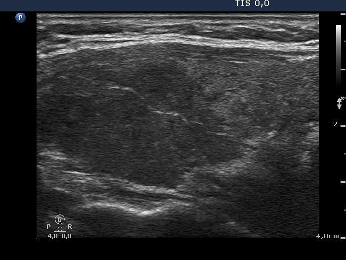
The sonographic pattern differs
significantly. There is a large, solitary, hypoechogenic nodule with
sharp borders in echonormal background in the left case while multiple
hypoechogenic lesions with irregular borders are found in a minimally
hypoechogenic background in the left case. The adenoma case present
signs of a type 1 vascular pattern and the vascularization is not
specific in the thyroiditis case.
|
|
Taking the sonographic and cytological data into account, we gave the
diagnosis of a follicular tumor.
|
Taking the sonographic and cytological
data into account, we gave the diagnosis of a lymphocytic thyroiditis.
|

