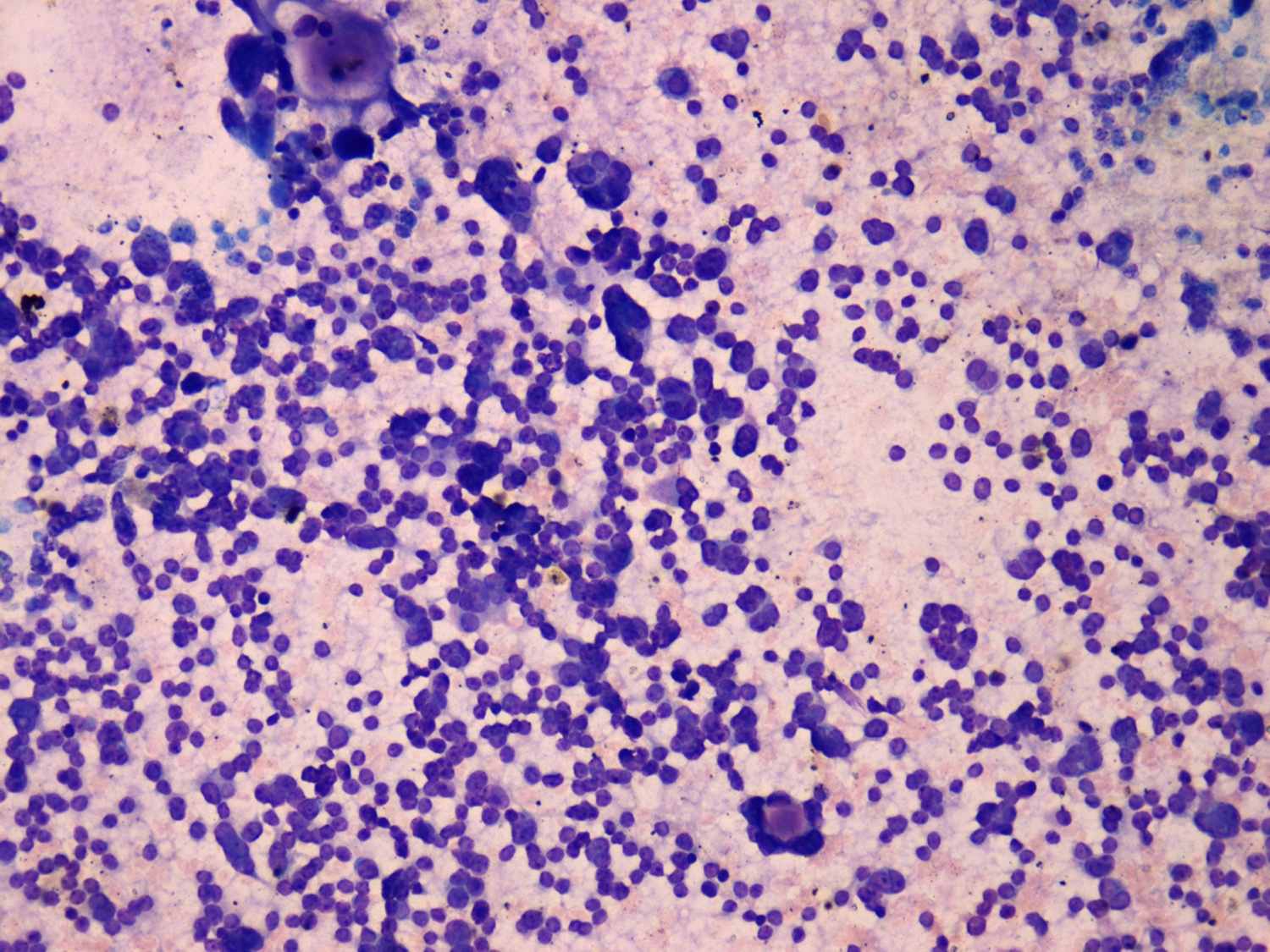
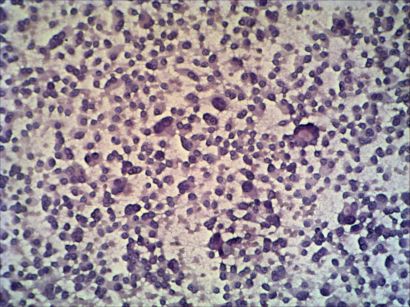
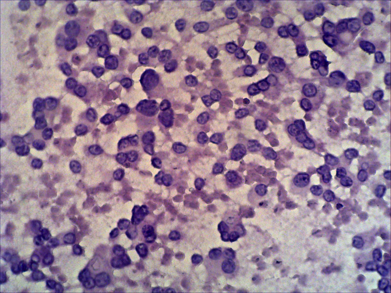
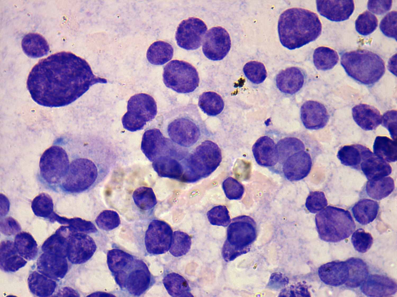
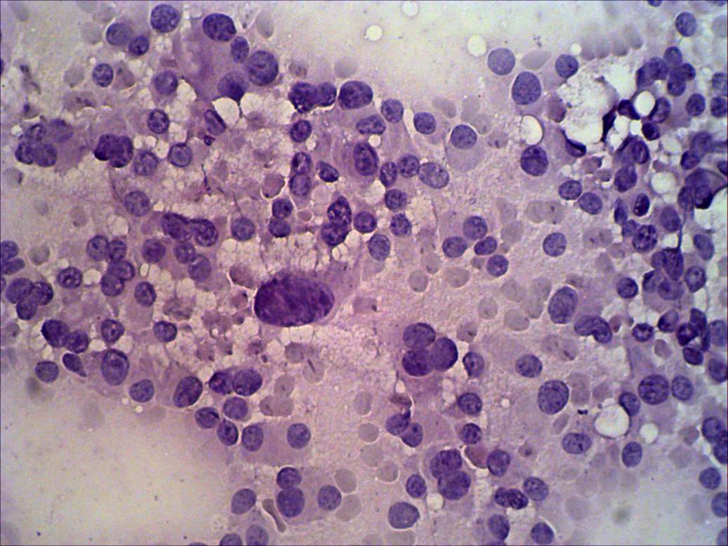
The cytological pattern of these two cases is very similar, almost
identical. Dispersed cells with abundant cytoplasm and eccentric nuclei
predominate the smear. In both cases a few enlarged, pleomorphic nuclei
can be
found. Some small cell groups resemble in both cases microfollicles.
The only difference is in the shape of the cells. Medullary carcinoma
cells are polygonal while those of Hürthle-cell adenoma are round.
Nevertheless, this difference is not enough for a clear
distinction. If the sonographic pattern is not pathognomic, serum
calcitonin determination is essential. |