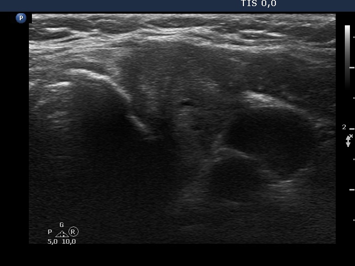
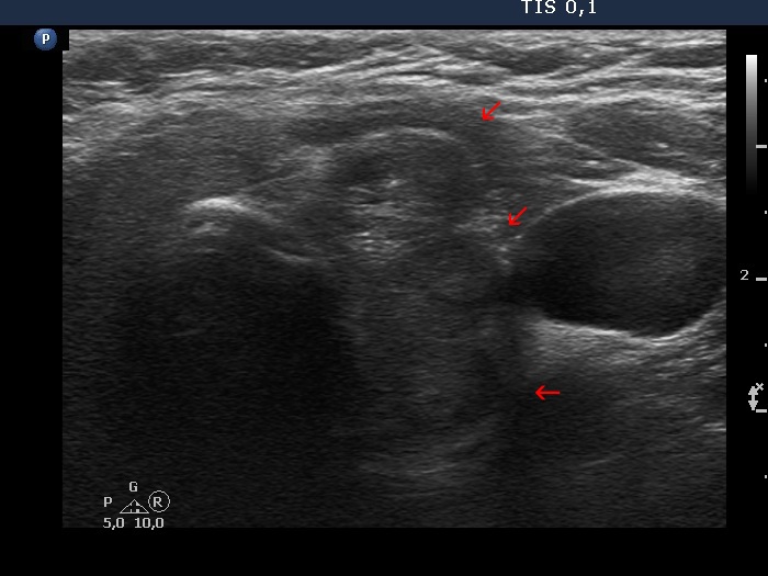
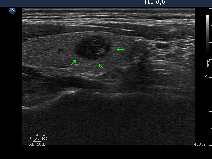
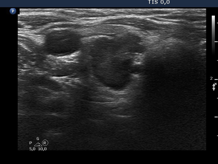
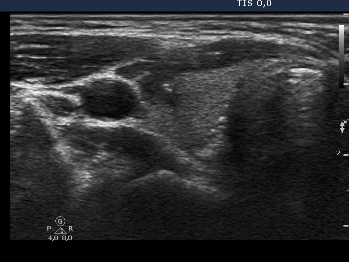
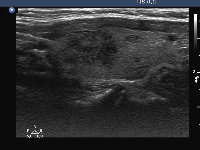
Benign, colloid goiter (cytology) - case 2110 |
Transverse scan |
Longitudinal scan |
 |
 |
 |
 |
This case illustrates the difference between pseudo-lobulation caused by the presence of multiple discrete lesions located next to each other and the true, pathognomonic lobulation. Red arrows point to different lesions causing a lobular appearance, while yellow arrows point to pathological protrusions in the margin of a single lesion.
|
|
|
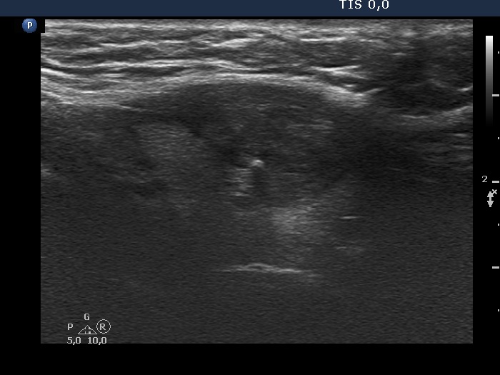
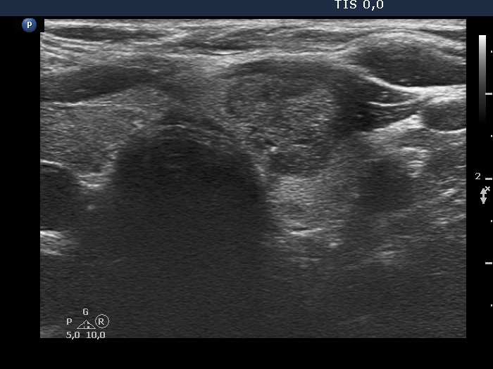
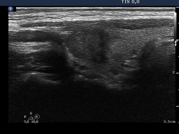
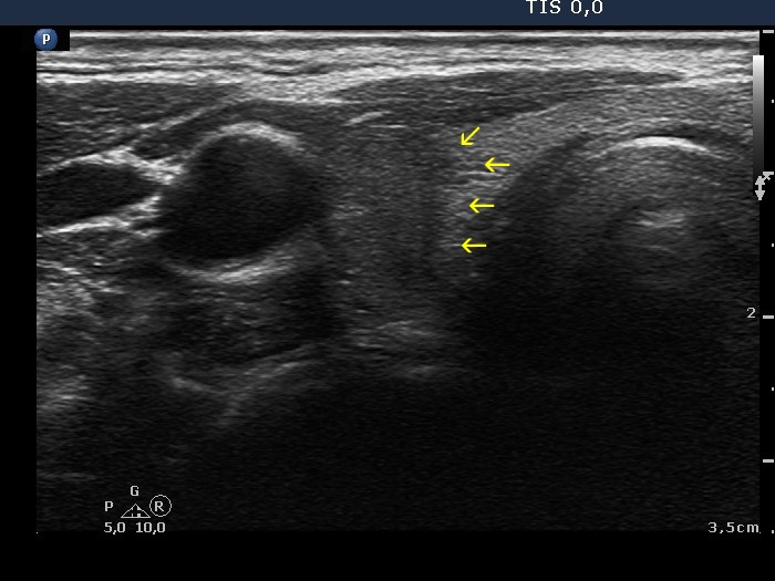
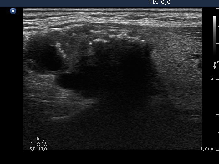
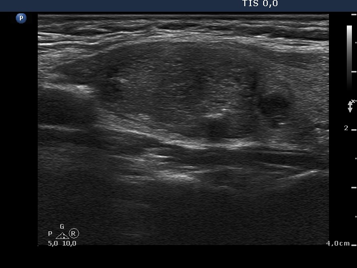
Benign, colloid goiter (cytology) - case 2083 |
Transverse scan |
Longitudinal scan |
 |
 |
 |
 |
Occasionally, it is difficult or even impossible to judge whether a lobulated surface is caused only by the presence of multiple discrete lesions or certain parts of a lesion are in fact pathologically lobulated. The clue is the thorough analysis of video. In this case, pathological lobulation is not likely.
|
|
|
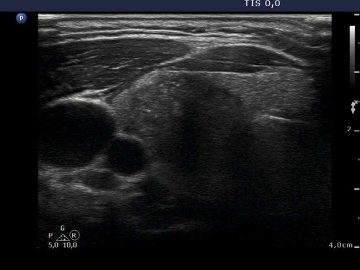
Benign, colloid goiter (cytology) - case 2154 |
Transverse scan |
Another transverse scan |
 |
 |
In this case, the lobulation is not pathological, this is caused by the presence of multiple lesions next to each other.
|
|
|
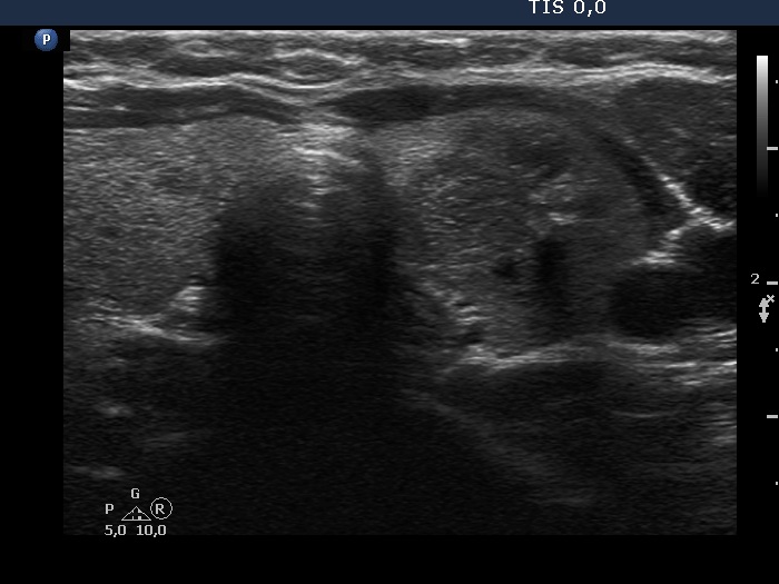
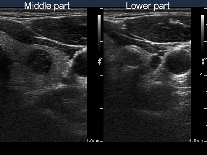
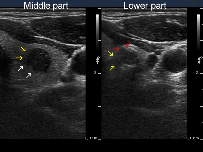
Papillary carcinoma (middle) and benign hyperplastic nodule (lower) (histology) - case conp034
|
Transverse scan |
Longitudinal scan |
 |
 |
 |
 |
The middle, malignant nodule has partly blurred (yellow arrows), partly lobulated margins (white arrows) while the lower, benign nodule presents blurred borders (yellow arrows) and spiculated margins (red arrows), as well. The small irregularities marked with green arrows cannot be judged as pathognomonic for lobulated margins.
|
|
|
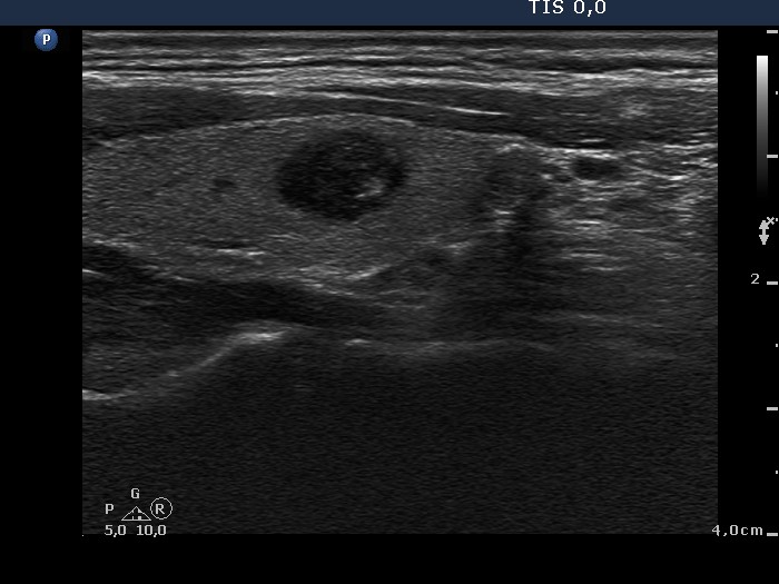
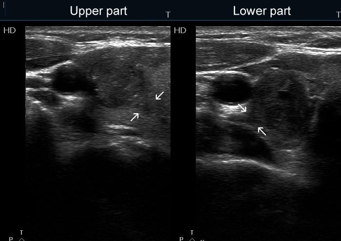
Benign, colloid goiter (cytology) - case 2121 |
Lower part of the right lobe, transverse scan |
Right lobe, longitudinal scan |
 |
 |
A large isoechoic lesion occupied the upper 3/4 of the right lobe. This lesion can be seen in the longitudinal scan. The hypoechoic nodule in the lower part presents typical lobulated margins.
|
|
|
|
Transverse scan |
Longitudinal scan |
 |
 |
 |
 |
The dorsal and medial borders of the nodule is partly ill-defined partly lobulated.
|
|
|
Papillary carcinoma (upper) and benign hyperplastic nodule (lower) (histology) - case conp027
|
Transverse scan |
Longitudinal scan |
 |
 |
 |
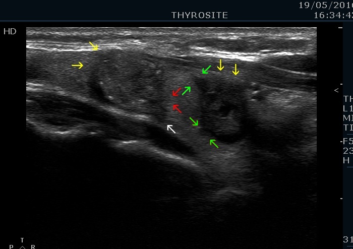 |
The nodules present different types of non-regular borders. White arrows point to spiculated margins, red arrows do to lobulated margins while yellow ones do to blurred borders. The origin of the spiculations on the surface of the hypoechogenic nodule (green arrows) is equivocal, these might be caused by the impression of the echonormal lesion on the surface. The upper, malignant nodule has microcalcifications while the lower benign one presents both taller-than-wide and longer-than wide sign.
|
|
|
|
Transverse scan |
Longitudinal scan |
 |
 |
 |
 |
In this case almost the entire border of the nodule is ill-defined, moreover the lesion has lobulated (yellow arrows) and spiculated (red arrows) margins, as well.
|
|
|
|
Transverse scan |
Longitudinal scan |
 |
 |
This nodule has clearly lobulated margins.
|
|
|
|
Transverse scans
|
 |
 |
The hypoechogenic tail marked with yellow arrows corresponds either to lobulated or spiculated margin.
|
|
|
|
Transverse scan |
Longitudinal scan |
 |
 |
This is a typical presentation of lobulated margins.
|
|
|
|
|
Transverse scan |
Longitudinal scan |
 |
 |
 |
 |
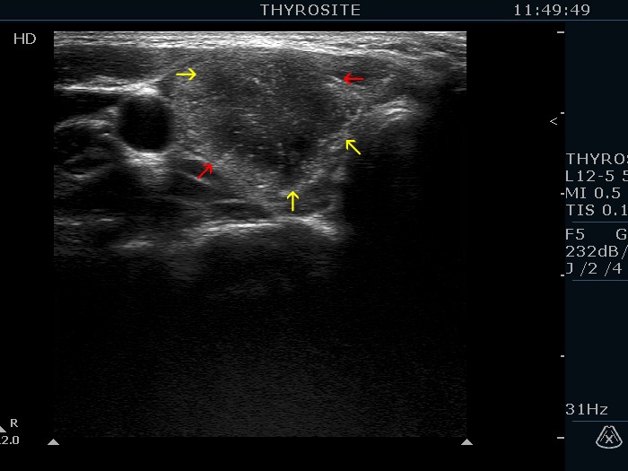
The border of the nodule presents small irregularities marked with red arrows. The interpretation of these small protrusions might be equivocal. There is one large clearly pathological lobulation marked with yellow arrow.
|
|
|
|
Transverse scan |
Longitudinal scan |
 |
 |
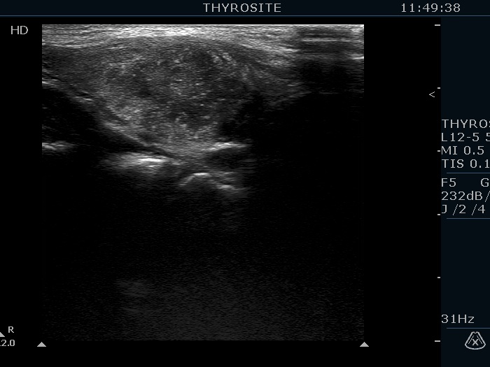
The interpretation of the borders is not evident in this case. The issue is whether this hypoechogenic lesion is one large heterogeneous nodule or is composed of multiple discrete nodules. In the former case, the nodule unequivocally has lobulated margins, while in the latter case the presence of pathological lobulations is doubtful. In my opinion, this hypoechogenic mass is more likely being one large nodule with heterogeneous echogenicity and therefore presents lobulated margins.
|
|
|
|
Transverse scan |
Longitudinal scan |
 |
 |
The nodule has partly blurred, partly lobulated margins.
|
|
|
|
Transverse scan |
Longitudinal scan |
 |
 |
The tumor presents lobulated margins.
|
|
|
|
Transverse scan |
Longitudinal scan |
 |
 |
The nodule has lobulated margins.
|
|
Subacute de Quervain's thyroiditis (cytology) - case 2169
|
Transverse scan |
Longitudinal scan |
 |
 |
The hypoechoic lesion has irregular spiculated, lobulated margins and presents blurred borders.
|
| |
|
