|
 |
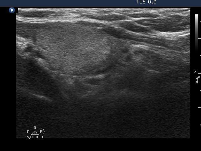 |
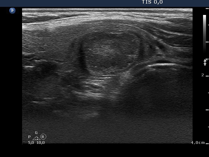
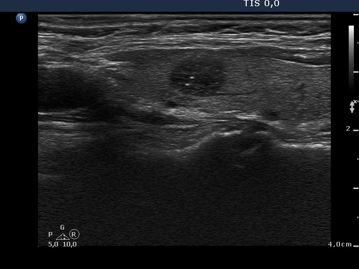
There is an isoechoic or a bit more echogenic nodule in the right lobe. We have to compare the echogenicity of the nodule to those parts of the lobe which are not influenced by technical influence. This is the ventromedial portion where the thyroid parenchyma is echonormal. The non-nodular thyroid dorsal to the nodule is moderately hypoechoic because the nodule' mass weakens the penetration of the ultrasound wave: thicker the nodule in the section, darker the non-nodular thyroid dorsal to the lesion.
|
|
Right lobe |
Left lobe |
 |
 |
 |
 |
Both nodules belong to the moderately hypoechoic subgroup because they are darker than the normal parenchyma but are brighter than the strap muscle.
|
| |
|
|
Right lobe |
Left lobe |
 |
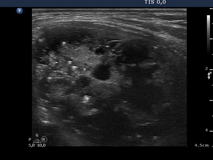 |
This dominantly solid nodule has isoechoic/echonormal solid area. Nevertheless, this is a highly suspicious nodule because of the presence of numerous microcalcifications.
|
| |
|
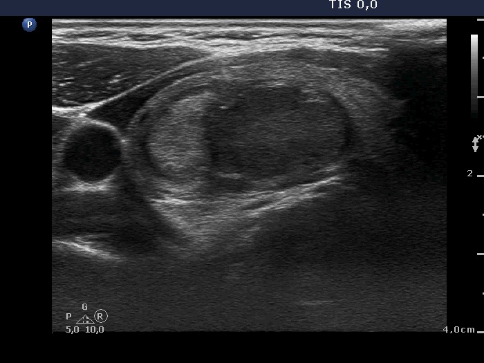
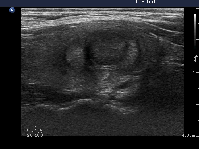
Benign hyperplastic nodules (histology) - case 1633 |
Transverse scan |
Longitudinal scan |
 |
 |
 |
 |
 |
 |
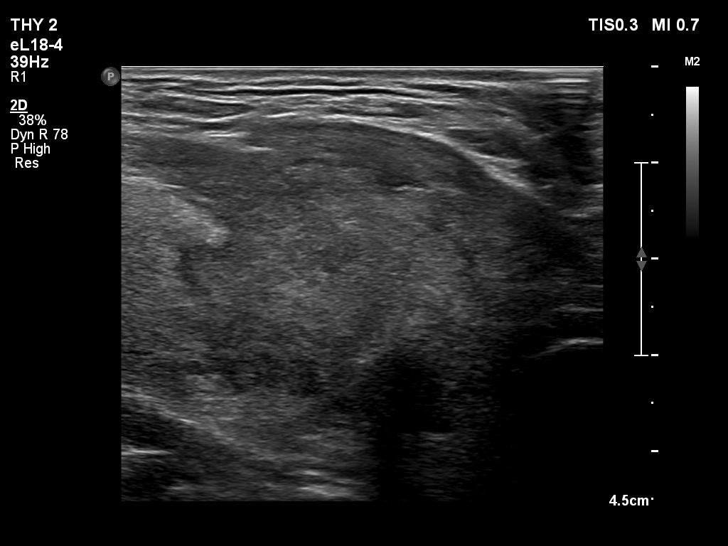
The lobe has multiple nodules, including hypoechoic and iso/hyperechoic lesions, as well. The hypoechoic nodule is brighter than the strap muscle, therefore this is a minimally/moderately hypoechoic lesion.
|
| |
|
Metastasis of a laryngeal carcinoma in the region of the thyroid (histology) - case 378 |
Right lobe |
Left lobe |
 |
 |
 |
 |
The right lobe (left images) has a moderately hypoechoic nodule while the left lobe (right images) has a deeply hypoechoic lesion.
|
| |
|
Parathyroid adenoma (histology) - case 671 |
Transverse scan |
Longitudinal scan |
 |
 |
 |
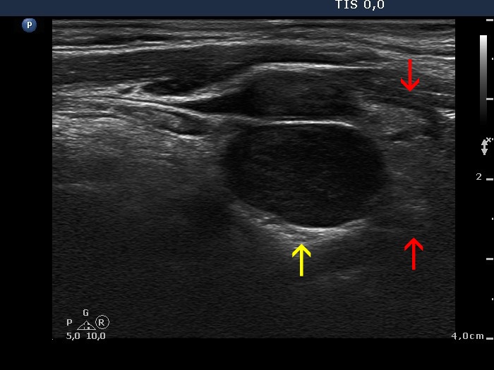 |
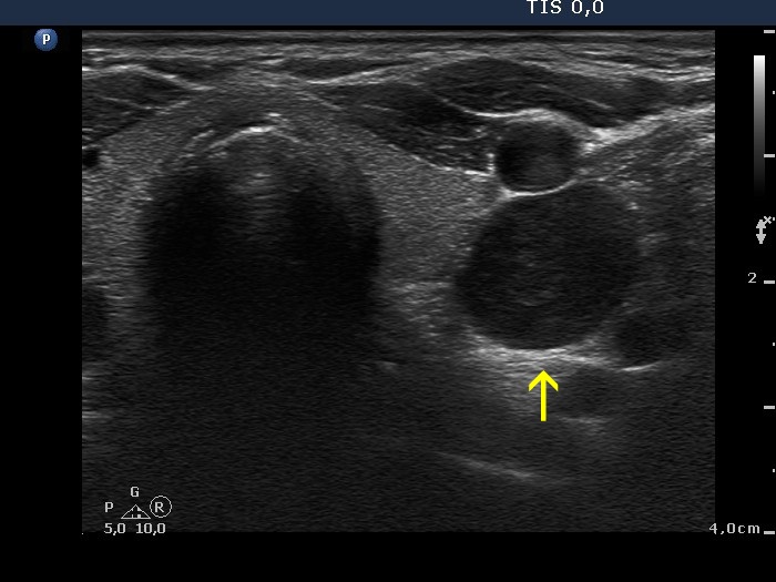
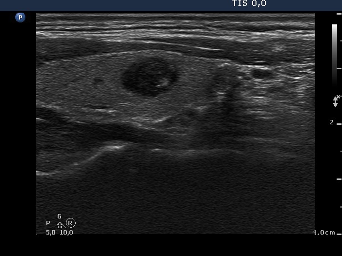
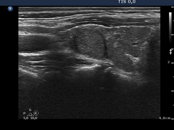
This is a deeply hypoechoic lesion which proved to be a parathyroid adenoma (yellow arrow). The longitudinal images prove that the adenoma was located upper to the thyroid (red arrows). Parathyroid adenomas are almost always hypoechoic.
|
| |
|
Parathyroid carcinoma (histology) - case 755 |
Transverse scan |
Longitudinal scan |
 |
 |
This is a hypoechoic lesion which proved to be a parathyroid carcinoma which was located in the lower pole of the left lobe. In the transverse view the tumor is brighter than the strap muscle while the situation is opposite in the longitudinal scan.
|
|
|
Oxyphilic adenoma (histology) - case 60 |
Transverse scan |
Longitudinal scan |
 |
 |
This is a deeply hypoechoic nodule because it is darker than the strap muscle.
|
| |
|
|
Right lobe |
Left lobe |
 |
 |
The echogenicity of the tumor in the right lobe is the same as that of the strap muscle. It means that this is a deeply hypoechoic nodule. The left lobe has a moderately hypoechoic nodule which proved to be benign on histopathology.
|
| |
|
|
Transverse scan |
Longitudinal scan |
 |
 |
The echogenicity of the nodule is same as that of the strap muscle. It means that this is a deeply hypoechoic nodule.
|
| |
|
Transverse scan |
Longitudinal scan |
 |
 |
This is a deeply hypoechoic nodule because it is darker than the strap muscle. Note the taller-than-wide shape and the irregular, lobulated margins.
|
| |
|
Benign nodule in Hashimoto's thyroiditis (cytology) - case 851 |
Transverse scan |
Longitudinal scan |
 |
 |
Depending on the definition of 'normal thyroid' the nodule can be considered either hyperechogenic or moderately hypoechoic, the reference tissue is the non-nodular parenchyma of the specific case or a healthy thyroid, respectively.
|
| |
|
|
Transverse scan |
Longitudinal scan |
 |
 |
The nodule is as hypoechoic as the strap muscle which means that this is a deeply hypoechoic lesion.
|
| |
|
 |
 |
The echo pattern of the nodule is almost identical to that of the extranodular part. Depending on the definition of 'normal thyroid' the nodule can be considered either isoechoic or moderately hypoechoic, the reference tissue is the non-nodular parenchyma of the specific case or a healthy thyroid, respectively.
|
| |
|
Follicular adenoma (histology) - case 500 |
 |
 |
The nodule is clearly more hypoechoic than the strap muscle running ventral to the thyroid. It means that this lesion is very or deeply hypoechoic.
|
| |
|
Metastasis of a malignant melanoma to the thyroid (histology) - case 1481 |
 |
 |
 |
 |
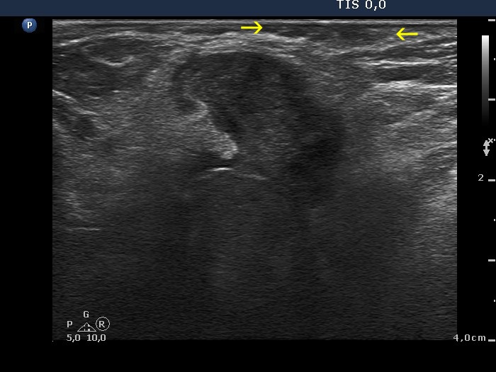
The nodule is clearly more hypoechoic than the strap muscle (marked with yellow arrows) running ventral to the thyroid. It means that this lesion is very or deeply hypoechoic.
|
| |
|
|
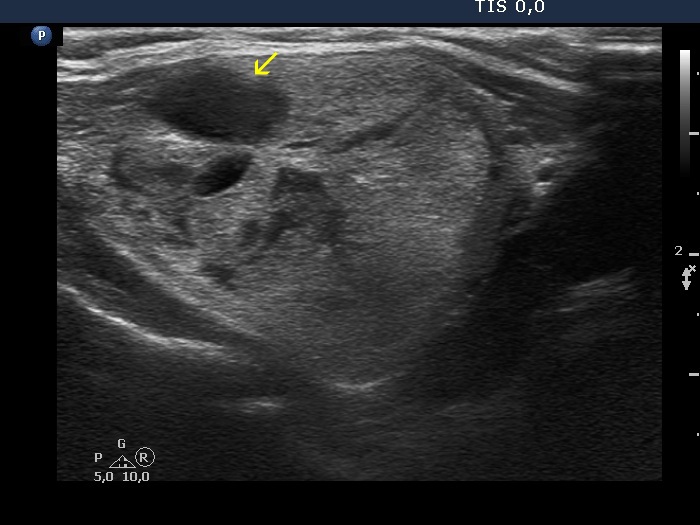
Right lobe, transverse scan |
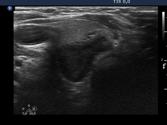
Right lobe, longitudinal scan |
 |
 |
 |
 |
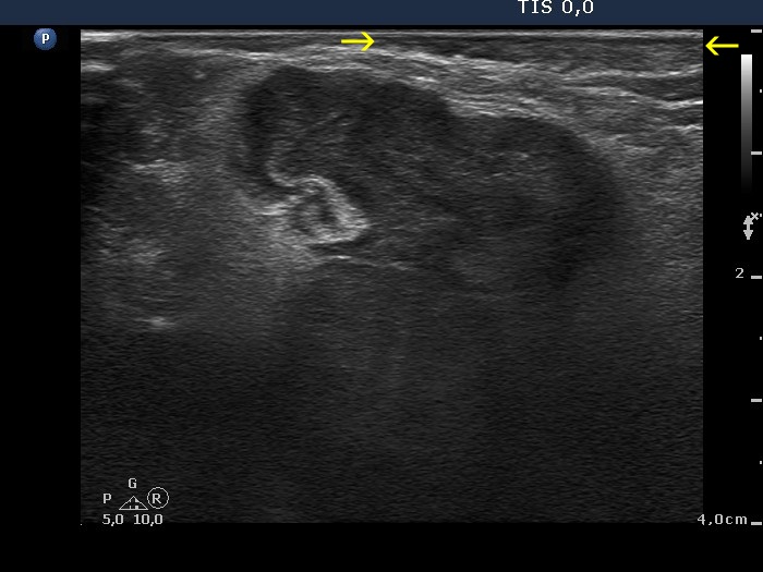
There are two nodules in the right lobe. The larger is echonormal or dominantly echonormal, heterogeneous which has cystic areas while the lower-dorsal is a deeply hypoechoic nodule. Regarding the former, the categorization depends on how to judge the ventral hypoechoic area marked with yellow arrow. If it we consider this cystic than the nodule is not heterogeneous, as the other hypoechoic areas neither reach 10% of the nodule nor are at least 10 mm in maximal diameter. If the area in question is considered solid than the nodule belongs to the dominantly echonormal, heterogeneous subgroup of nodules.
|
|
