|
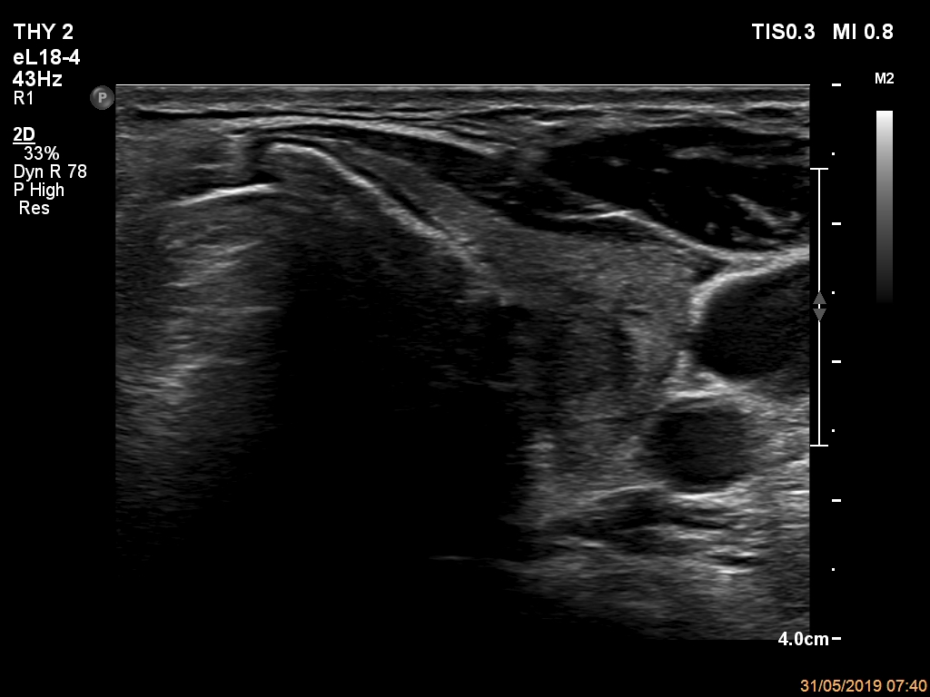
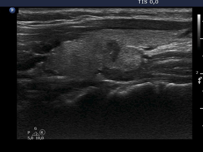
Transverse scan
|
 |
 |
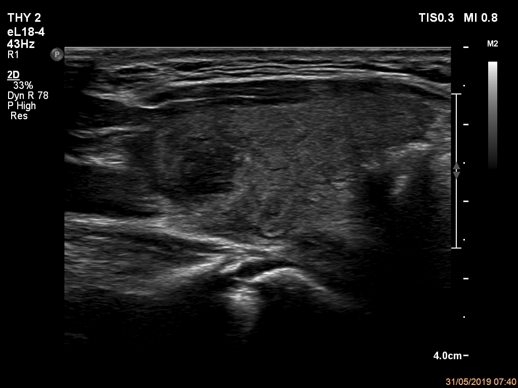
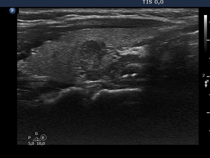
Longitudinal scan
|
 |
 |
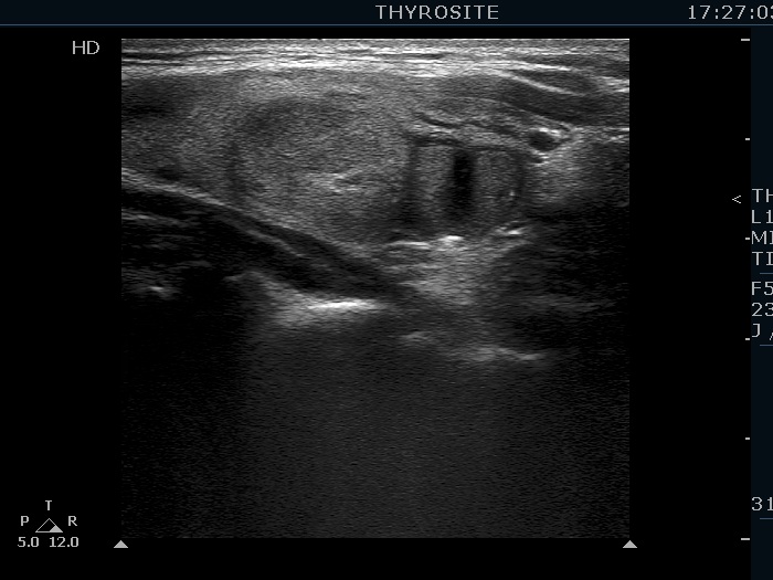
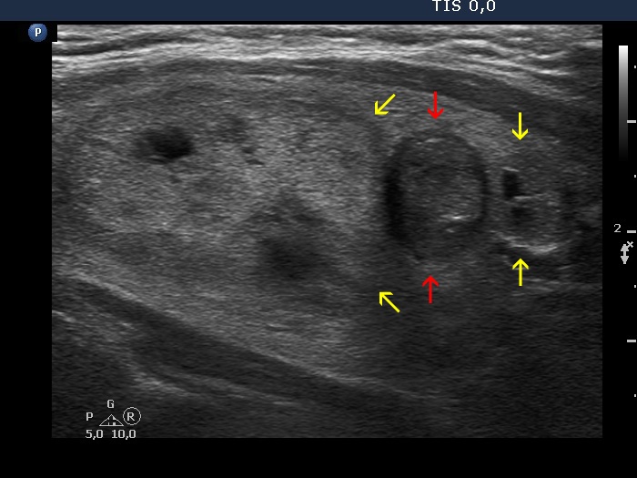
This nodular area presents nonparallel orientation in the upper right image which shows the lower part of the nodule. In this location the thyroid is normally narrowed, and the ratio of width to depth of the normal gland is usually < 1. Therefore, a nodule located in the lower pole can present non-parallel orientation simply because it follows the shape of the lobe. It is worth viewing the video.
|
| |
|
 |
 |
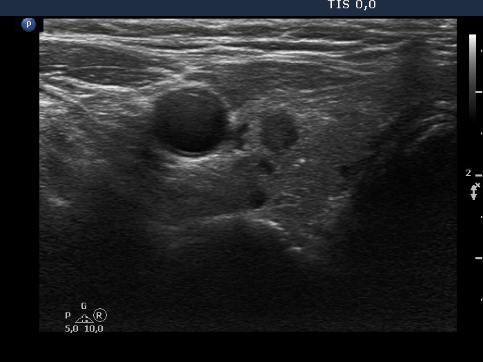
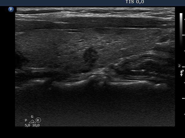
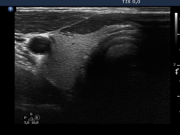
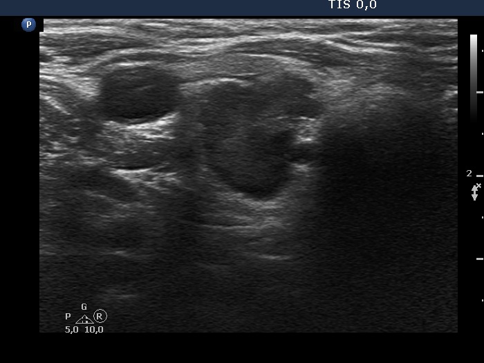
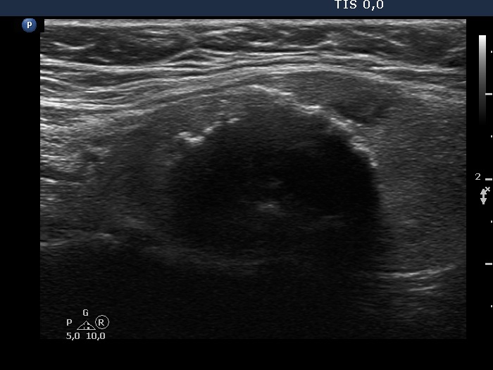
The small nodule shows irregular shape, but this is not of pathological importance. The hard wall of the carotid artery prevents the nodule from spreading sideways.
|
| |
|
Hashimoto's thyroiditis (histology) - case 1096 |
 |
 |
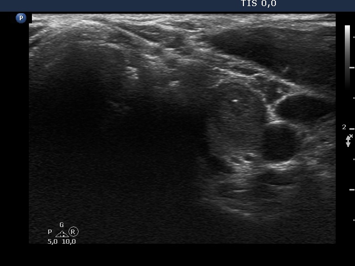
The lesion in the dorsal part of the lobe shows taller-than-wide shape. However, this is not a pathological form of nonparallel orientation. The taller-than-wide sign is caused by the anatomy, the trachea and the carotid artery hinder the growth of the lesion sideways. Moreover, the lesion proved to be not a true nodule on histopathology.
|
| |
|
|
Right lobe, transverse scan |
Left lobe, transverse scan |
 |
 |
Both lobes present taller-than-wide shape. In such lobes a nodule simply follows the shape of the lobe.
|
| |
|
|
Upper part of the right lobe, transverse scan |
Middle-lower part of the right lobe, transverse scan |
 |
 |
The upper nodule shows taller-than-wide shape similarly to that part of the lobe, while the nodule in the lower two-third of the lobe does regular, parallel orientation.
|
| |
|
|
 |
 |
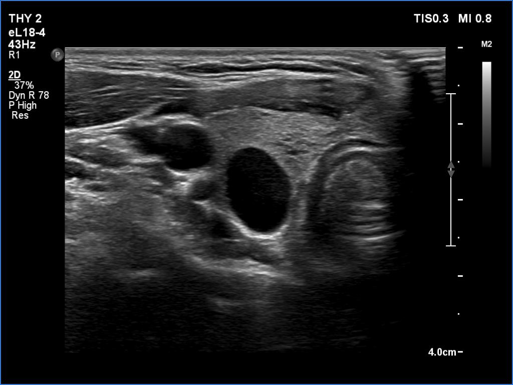
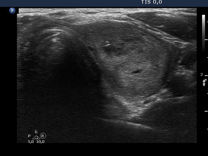
The nodule shows taller-than-long shape. However, the nonparallel orientation is caused by the anatomical situation: the lesion was sandwiched between two other nodules. The latter are signed with yellow arrows.
|
| |
|
|
 |
 |
 |
 |
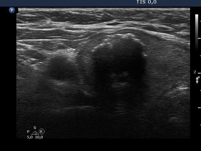
Four images gained on longitudinal scan are presented. The issue is the same as in the previous case: is it a discrete area composed of a single or multiple nodules. In my opinion, it is rather a single nodule: we can see on the left lower image an irregularly shaped echonormal area, the tail of which can be found even in the hypoechogenic part of the lesion.
|
| |
|
|
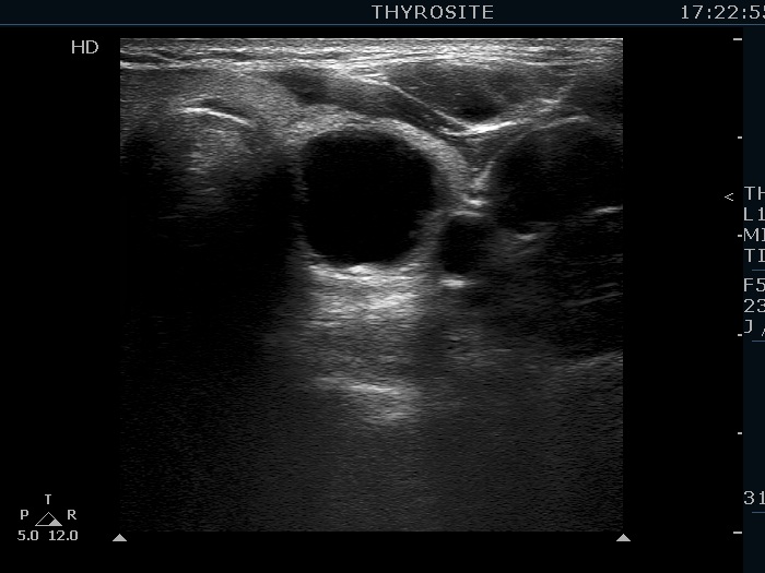
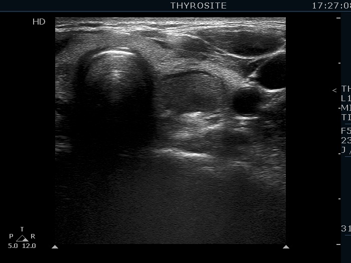
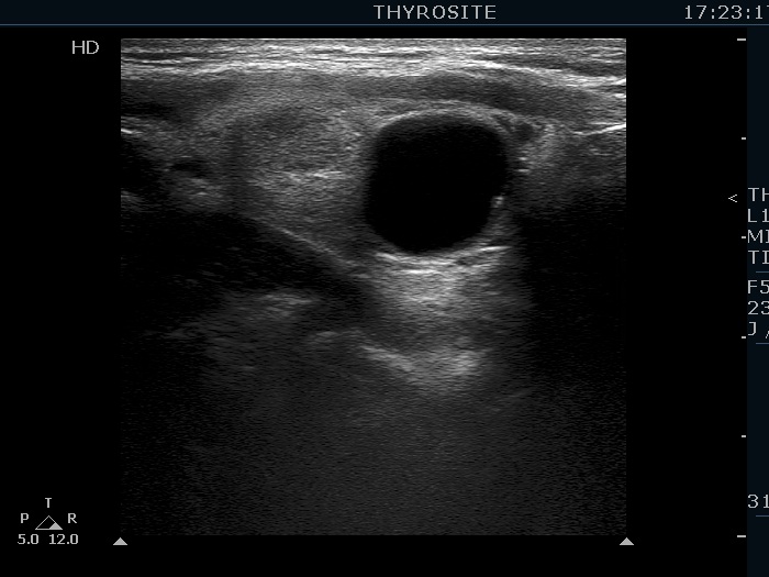
Upper part of the right lobe, transverse scan |
Middle-lower part of the right lobe, transverse scan |
 |
 |
Upper part of the right lobe, longitudinal scan
|
Lower part of the right lobe, longitudinal scan
|
 |
 |
The shape of the small hypoechoic nodule is close to taller-than-wide in transverse scan (right upper image) while clearly shows taller-than-long shape on longitudinal sections (right lower image). However, the latter is caused by the large nodule which hinders the normal growth in the upway direction.
|
| |
|
Benign lesion (cytology) |
|
Transverse scan |
 |
 |
Longitudinal scan
|
 |
 |
Although at first sight, the nodule seems to show taller-than-wide shape and both taller-than-wide and taller-than-long shape, left and right case, respectively. Indeed, the acoustic shadow caused by the macrocalcification makes the judgement of the nodule' depth impossible.
|