Selected topics - intranodular hyperechogenic figures - Table 4 (large). Punctate echogenic foci (including microcalcifications) |
||
A punctate echogenic focus is a round bright granule. The size of this figure is less than 1 mm, usually is around 0.5 mm. A lesion which has typical punctate echogenic foci (microcalcifications) almost always presents less bright granules, too. The latter might be non-typical forms of microcalcifications or non-specific granulations. In fact the identification of punctate echogenic foci (including microcalcifications) is a matter of exclusion other forms of hyperechogenic figures, i..e comet-tail artifacts, connective tissue and posterior back wall enhancement. All of the latter have additional features lacking in the event of punctate echogenic foci. Nevertheless, it means that atypical presentations of these figures might mimic punctate echogenic foci including microcalcifications.
The presentation of punctate echogenic foci depends highly on the ultrasound device. If the resolution is worse than the granule is larger (see the lowest image). On the other hand there is a very harmful trend in newer equipments which might cause difficulty in recognition of punctate echogenic foci (microcalcifications). This trend is the "overharmonization" which means that the normal grainy structure of the thyroid is masked in order to gain a more harmonic, beautiful image. It has practically no sense, moreover the overharmonization masks the pathologic hyperechogenic granules, the microcalcifications, too, which is a very serious hazard.
We prefer the term punctate echogenic foci instead of microcalcification in order to avoid misnomer for granules caused by other pathological structures. Nevertheless, in many publications and even in other parts of this website we use the term "microcalcification" for every punctate echogenic foci.
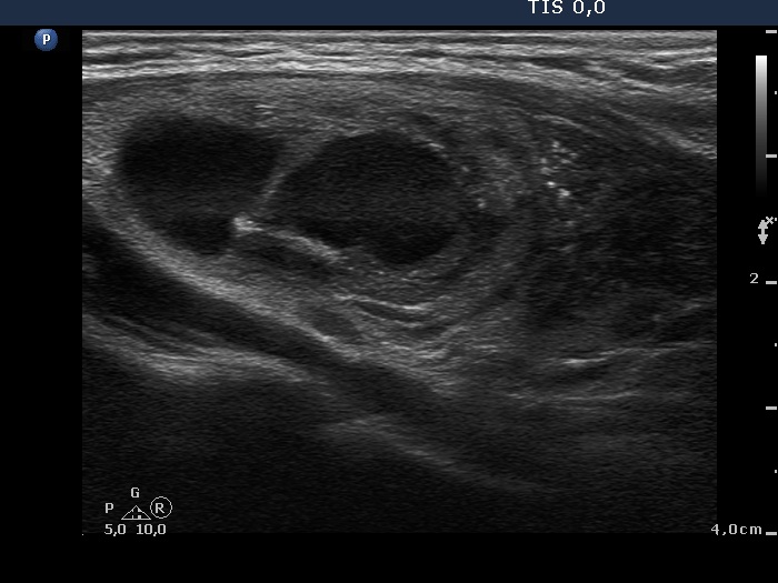
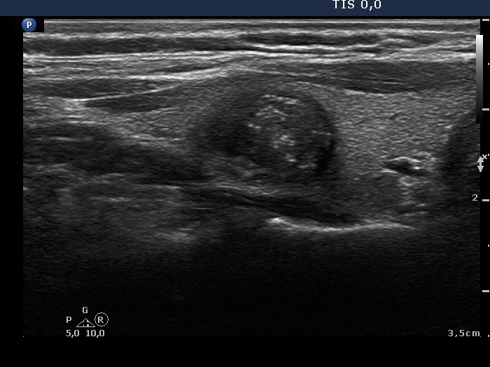
Papillary carcinoma (histological diagnosis) - case 763 |
|
 |
 |
There are at least 6 bright granules in the left while in the right image. The lesion as most nodule contains non-specific granules and lines which are much less bright than the former. The granules of intermediate brightness might be punctate echogenic foci, as well. |
|
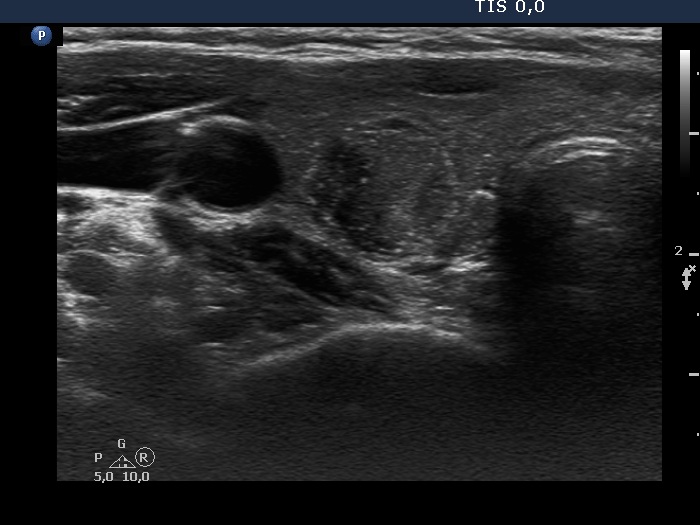
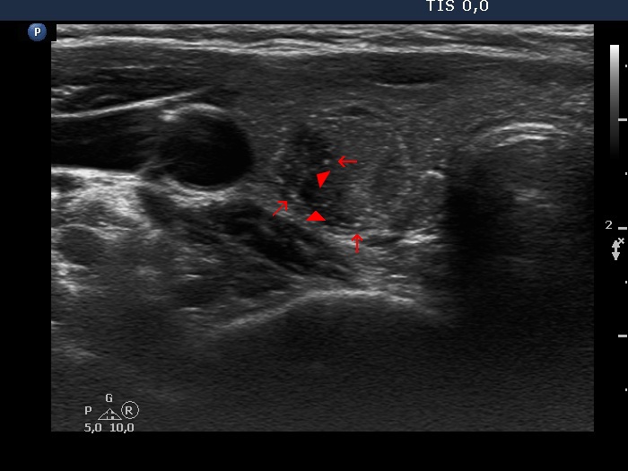
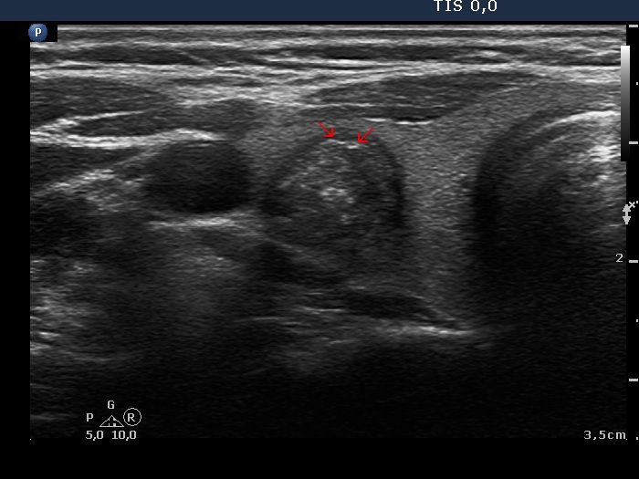
Papillary carcinoma (histological diagnosis) |
|
 |
|
 |
 |
This case is less edifying or may be more edifying. Compared with the previous case, the granules here are less bright. Nevertheless, great proportion of them belong to punctate echogenic foci (arrows). It is worth to compare these with non-specific granules (arrowheads). |
|
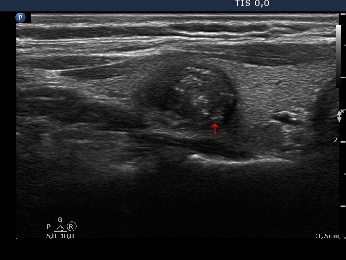
Papillary carcinoma (histological diagnosis) - case 607 |
|
 |
 |
 |
 |
Arrowheads point to several non-specific granules while arrows do to punctate echogenic foci. |
|
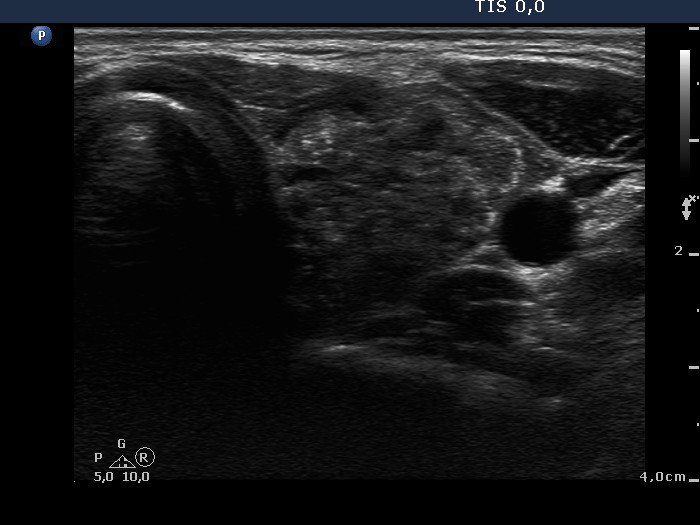
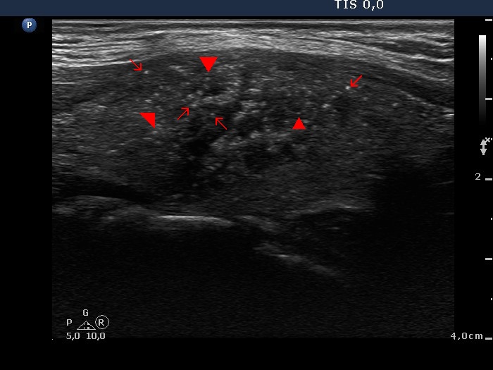
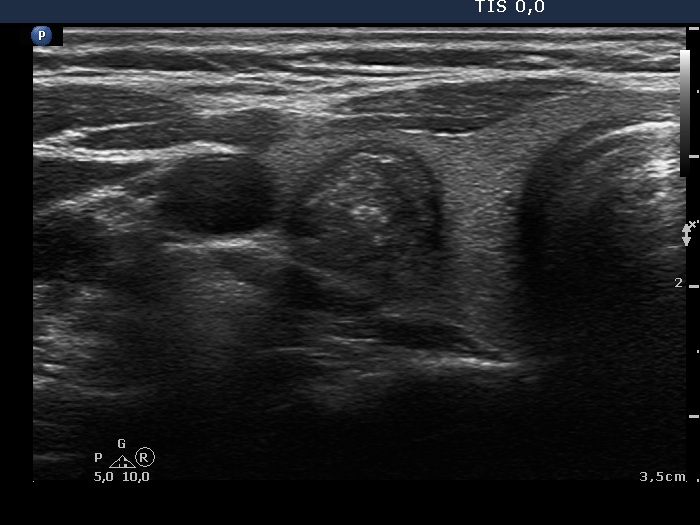
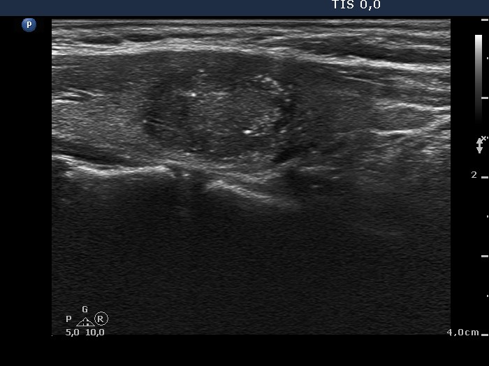
Benign hyperplastic nodule (histological diagnosis) |
|
Upper and lower horizontal views |
Longitudinal view |
  |
 |
The bright granules cannot be categorized other than punctate echogenic foci (microcalcifications). The presentation in the right image is very close to the starry sky phenomenon. Fortunately, such pattern is very rarely seen in benign lesions. |
|
Benign cystic-colloid goiter (cytological diagnosis) |
|
 |
 |
 |
 |
This is an ambiguous pattern: although hyperechogenic bright granules predominate the lesion, a few similarly bright figure seems to be linear (arrows). On the other hand two of them are in fact figures composed of two granules (in the left image). |
|
Papillary carcinoma (histological diagnosis) - case p061 |
|
 |
 |
There is only one granule suspicious being an punctate echogenic focus (microcalcification) in the left, horizontal view while the right, longitudinal view has more typical forms. |
|
Papillary carcinoma (histological diagnosis) - case 469 |
|
 |
 |
This tumor contained numerous punctate echogenic foci (microcalcifications). |
|
Hashimoto's thyroiditis (cytological diagnosis) - case 1137 |
|
 |
 |
A solitary punctate echogenic focus is presented in the right image. |
|
Papillary carcinoma (histological diagnosis) - case p009 |
|
 |
 |
On histopathological analysis numerous psammoma bodies were found in this tumor. On the other hand the ultrasound presentation is not very convincing. This case illustrates that such pale granules might also be microcalcifications. |
|
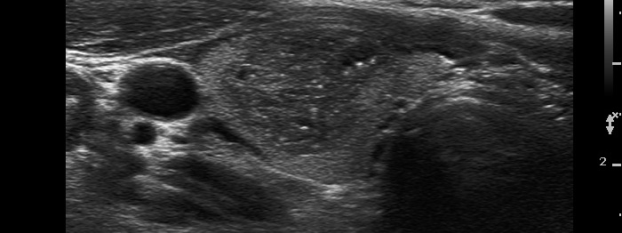
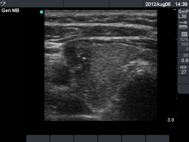
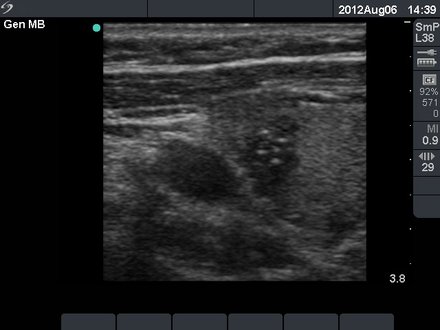
Papillary carcinoma (histological diagnosis) - case 882 |
|
 |
 |
Smaller and larger bright granules correspond to punctate echogenic foci, in this case to microcalcifications. |
|
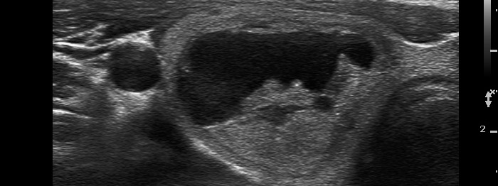
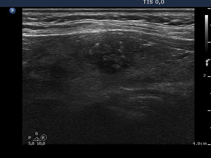
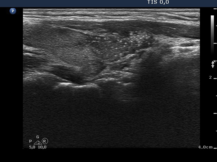
Papillary carcinoma (histological diagnosis) - case 684 |
|
 |
 |
This investigation was performed with an older equipment with worse resolution, therefore the granules are not only larger but a bit blurred compared with punctate echogenic foci presented in the former cases. |
|