Benign nodular hyperplasia - Case 40. (ultrasonographic picture 5)
|
|

|
|
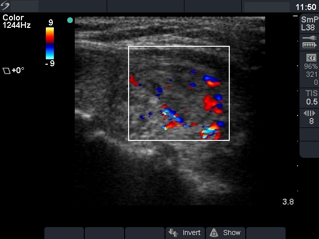
Other section of the left lobe, longitudinal scan, color Doppler mode. Both peri- and intranodular blood flow can be seen.

|
|
|
|
Other section of the left lobe, longitudinal scan, color Doppler mode. Both peri- and intranodular blood flow can be seen.