
|
|
Chronic lymphocytic thyroiditis - Case 58.
|

|


Clinical data: a 45-year-old woman requested a second opinion. Cytology resulted in the 'suspicion of thyroid cancer' in another hospital.
Palpation: both lobes were firm and their surface was irregular.
Functional state: euthyroidism with TSH-level 1.11 mIU/L. Anti-TPO below 10 IU/mL.
Scintigraphy: indicated a 'cold' nodule in the right lobe.
Ultrasonography: there was a hypoechogenic thyroid with several circumscribed areas. We aspirated a nodule-like lesion with increased intranodular blood flow in the right lobe. (It corresponded to the 'cold nodule'.)
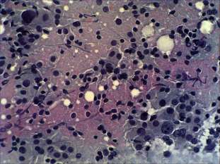
Cytological picture: there was diffuse colloid precipitate in the background. Follicular cells were arranged in monolayered sheets and in small clusters. They varied in size, part of them showed oxyphilic metaplasia. There were inflammatory cells, epitheloid cells and nuclear debris in the background. A few multinucleated giant cells were also present.
Combined ultrasonographic-cytological diagnosis: benign Hashimoto's thyroiditis.
The patient underwent surgery because of the result of first FNAC.
Histopathology: Hashimoto's thyroiditis without any nodule.
Comment: the tool for diagnosing a nodule has to be not the palpation and not the palpation-based scintigraphy, but ultrasonography. This patient had no nodule on ultrasonography, therefore the possibility of a papillary cancer was only theoretical. Nevertheless, the cytologic picture was not very reassuring.


