
|
|
Chronic lymphocytic thyroiditis - Case 59.
|
|


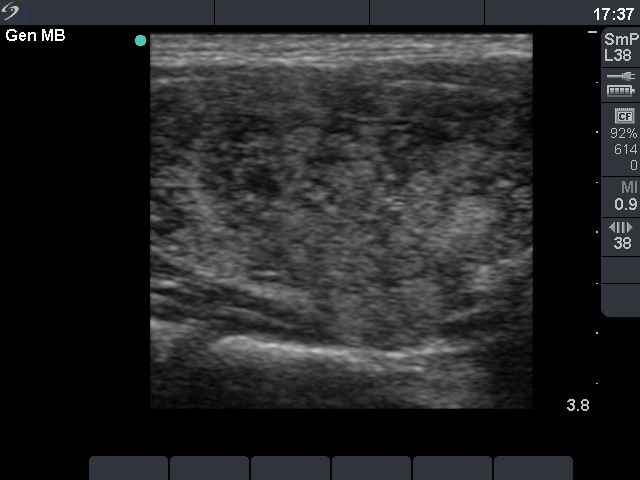
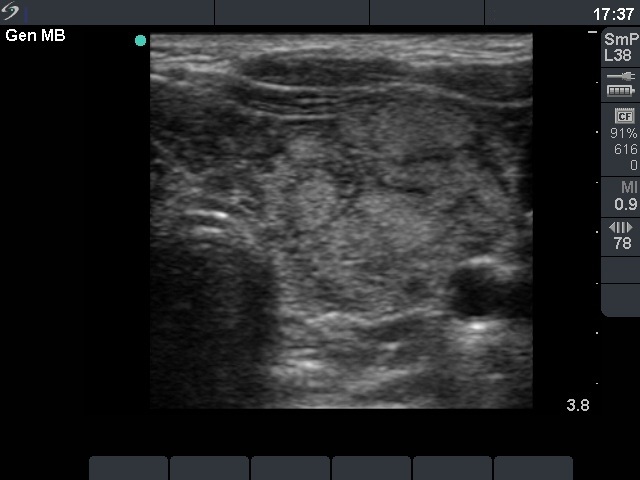


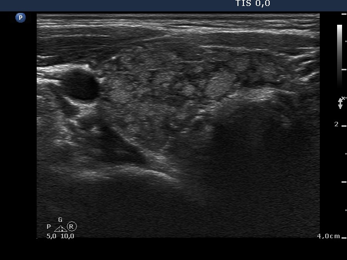
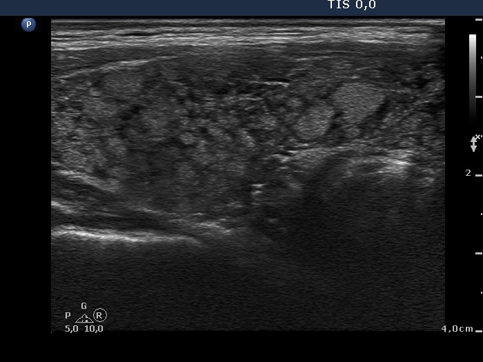


First examination (first and second rows of images):
Clinical presentation: a 25-year-old woman was referred for evaluation of infertility. Both thyroid lobes were firm on palpation.
Functional state: hypothyroidism with TSH 11.6 mIU/L.
Ultrasonography revealed a typical example of the so-called micronodular form of Hashimoto's thyroiditis. It means that the thyroid is composed of numerous echonormal nodule-like lesions with a maximal diameter of 5 mm to 15 mm. These look like secondary and tertiary lobules of the lobe in reality. The 'extranodular' parenchyma was hypoechogenic.
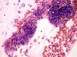
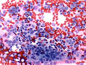
FNAC: disclosed lymphocytic thyroiditis.
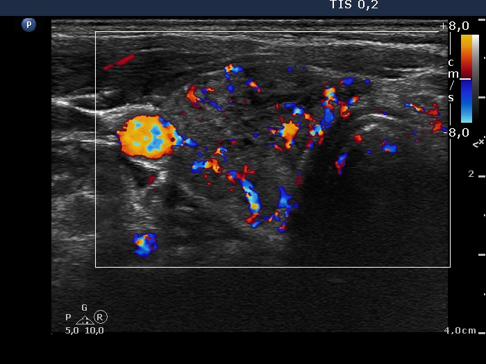
Follow-up examination 5 years later (third row of images):
Clinical presentation: the patient had no complaints she was referred for follow-up examination.
Functional state: euthyroidism on daily 75 microgram levo-tiroxine (TSH 1.83 mIU/L).
Ultrasonography: the size, the echo pattern of the thyroid, i.e. the echogenicity index, the vascularization were almost identical to that seen for 3 years.
Suggestion TSH-test every year.