
|
|
Chronic lymphocytic thyroiditis - Case 60.
|
|


Clinical data: a 19-year-old woman was referred to another hospital for the evaluation of weight loss and palpitation. Ultrasonography revealed multiple 'nodules'. The endocrinologist suggested FNAC.
Palpation: no abnormality.
Functional state: euthyroidism (TSH-level 0.99 mIU/L).
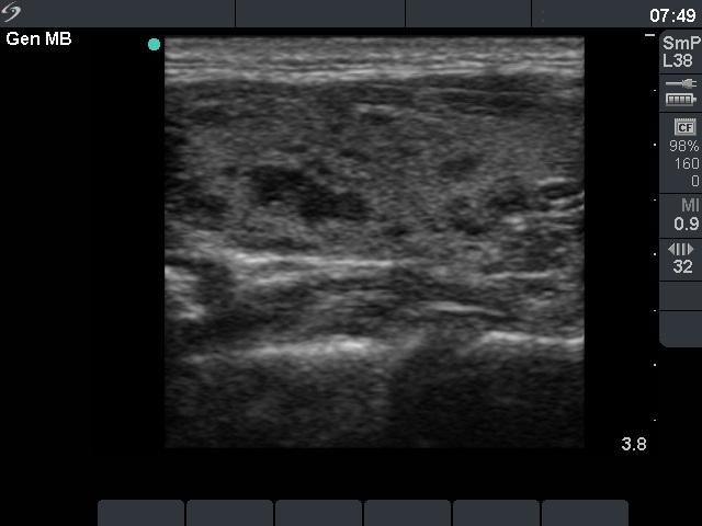
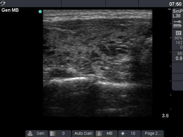
Ultrasonography: there were multiple small hypoechogenic areas in both lobes. The borders of these lesions were irregular. The US pattern corresponded to autoimmune thyroiditis.
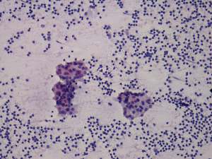
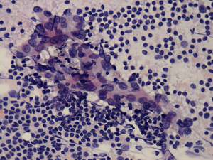
Cytological picture: there is no colloid in the background. Clusters of follicular cells were in heterogeneous lymphoid background.
Cytological diagnosis: Hashimoto's thyroiditis. Anti-TPO was also performed and was in the normal range (8 U/mL).
Suggestion: regular follow-up investigations.
A year later the patient became pregnant. During the 6th week of the pregnancy the TSH resulted in 6.9 mIU/L. Levo-tiroxine was administered.
Comment: the greatest issue in thyroid ultrasonography is the misinterpretation of hypoechogenic areas of Hashimoto's thyroiditis as nodules. Although the US pattern was unequivocal, we performed FNAC mainly for a psychological reasons. The term 'nodule' caused great fear of cancer. In this case, there was a second reason of aspiration: the negative anti-TPO test.