
Papillary carcinoma - Case 9. (ultrasound picture 4)
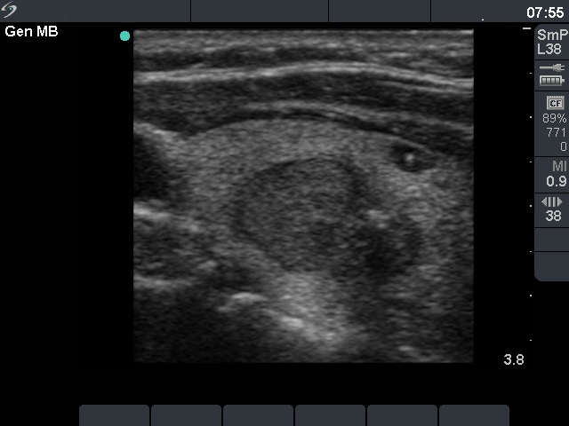 Left lobe, longitudinal view. Two nodules next two each other, the upper (left on the image) proved to be an oxyphilic adenoma while the lower (right on the image) did a papillary microcancer. The smallest lesion located in the ventral part proved to be a hyperplastic small nodule on histopathology. |
