
|
|
Papillary carcinoma - Case 20.
|

|






Clinical data: a 31-year-old woman was evaluated in another hospital because of a nodular goiter 2 years earlier. Aspiration cytology resulted in a benign lesion. She was referred for a follow-up examination. She had no complaints.
Palpation: a hard nodule in the isthmic part of the right lobe.
Functional state: euthyroidism (TSH-level 0.61 mIU/L).
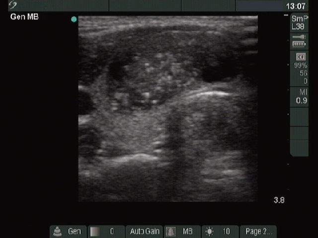
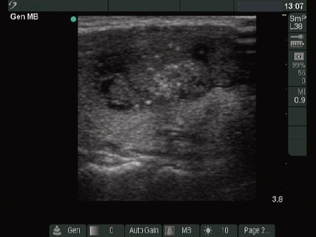
Ultrasonography: there was a hypoechogenic nodule in the isthmic part of the right thyroid. The nodule contained hyperechogenic foci of various size, part of them corresponded to microcalcifications. The larger ones resembled that frequently observed in the case of medullary cancer. The intranodular blood flow was increased. Only one diameter of the nodule was given in report of former ultrasound, it was 25 mm. On the present examination, the dimensions of the nodule were 29x15x36 mm.
Cytology: first aspiration cytology was not diagnostic. We performed serum calcitonin test which resulted in normal value (below 0.59 pM/L). We repeated the FNAC.
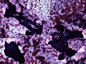
Cytological diagnosis: suspicion of papillary cancer.
Histopathology: papillary carcinoma.


