
|
|
Intranodular hyperechogenic figures - case 1425
|
|

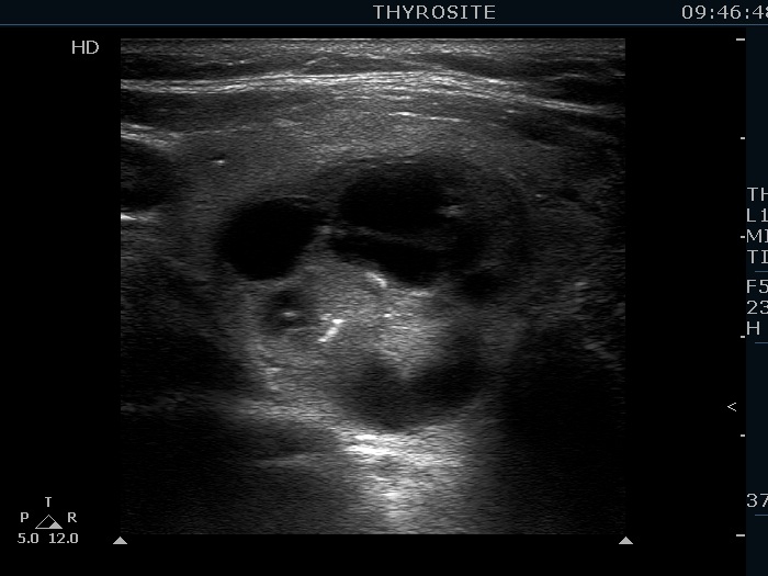

Clinical presentation: a 64-year-old woman requested evaluation of 'lump in the throat' feeling. We examined her 8 years ago when a multinodular goiter was found and cytology was benign.
Palpation: a multinodular the right lobe.
Functional state: euthyroidism with TSH 0.29 mIU/L, FT4 15.1 pM/L.
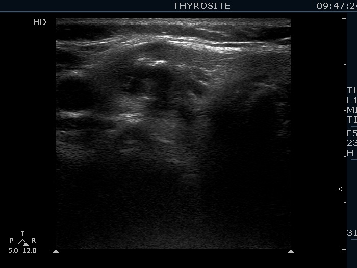
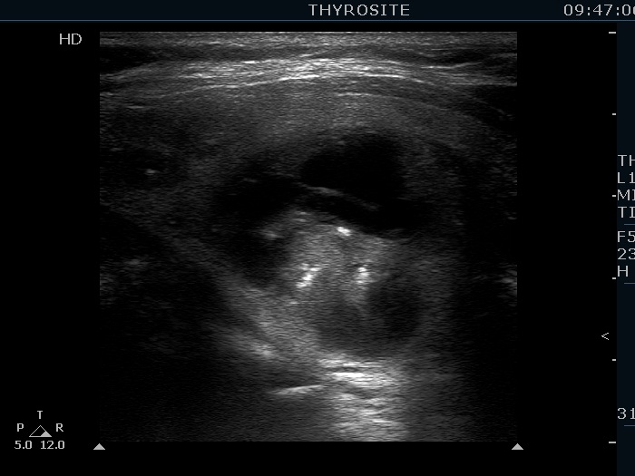
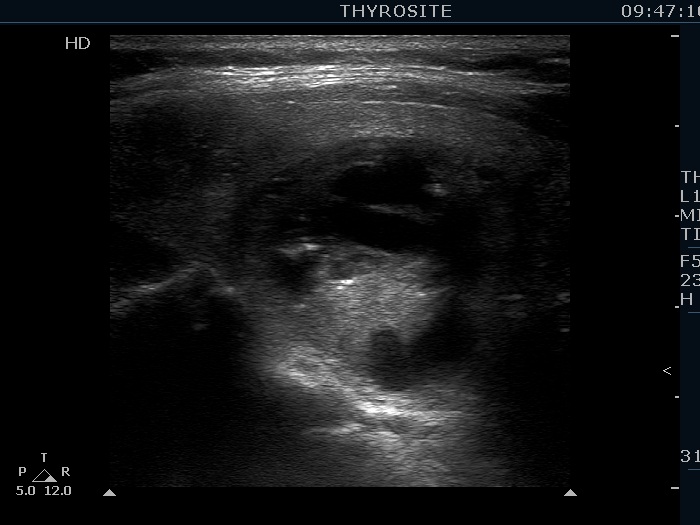
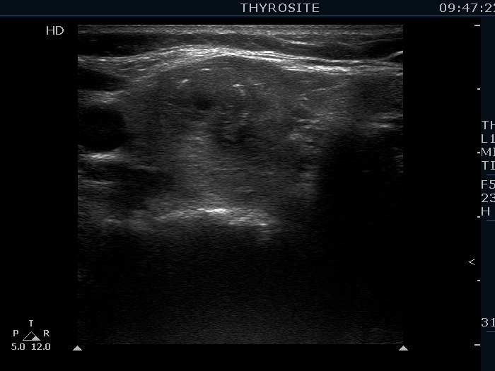
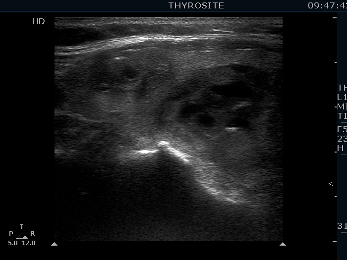
Ultrasonography. The thyroid was echonormal. There were two lesions in the right lobe, the upper was a minimally hypoechogenic and had bright hyperechogenic figures and non-.specific granules. The lower lesion was a multi-chambered cyst with echonormal solid area. This nodule contained larger linear and granular hyperechogenic figures corresponding to connective tissue. None of the nodule has increased since the first visit.
Comment.
-
Although the bright hyperechogenic granules in the upper nodule might correspond to microcalcifications, the ultrasound presentation of the nodule is not suspicious.
-
The thick hyperechogenic figures in the lower, larger nodule are relatively unusually presentations of connective tissue.