
|
|
Intranodular hyperechogenic figures - case 155
|
|

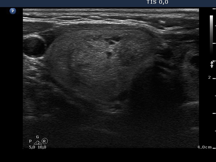

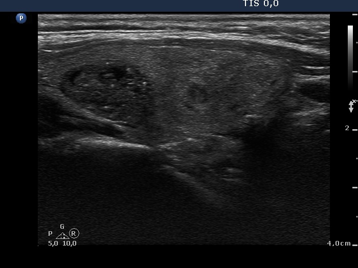

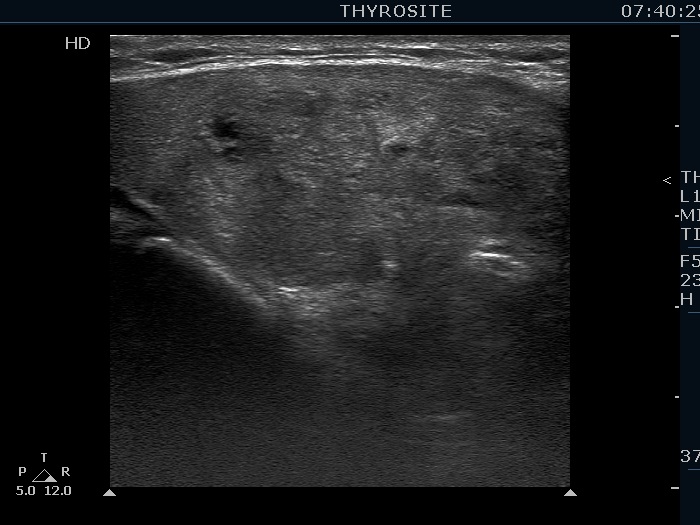


First examination (first and third rows of images):
Clinical presentation: a 57-year-old woman was referred for aspiration cytology of a multinodular goiter has been known for more than two decades. The patient had no complaints.
Palpation: a multinodular goiter without any nodule suspicious on palpation.
Functional state: euthyroidism with TSH 0.96 mIU/L.
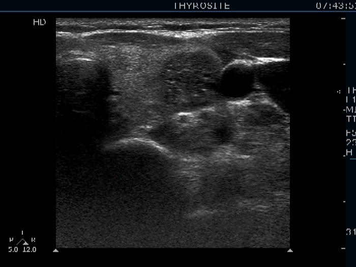
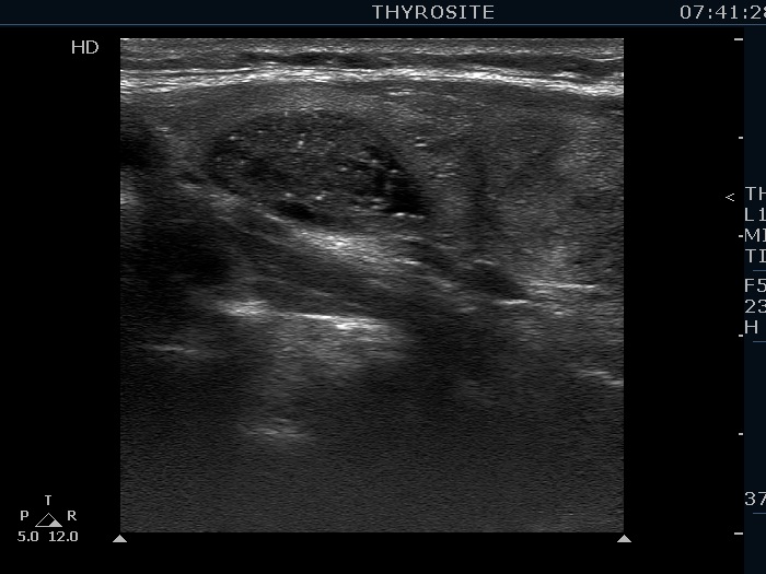
Ultrasonography. The thyroid was echonormal and was composed of multiple nodules. Most of them were hyperechogenic or minimally hypoechogenic. There was a hypoechogenic nodule presenting bright hyperechogenic granules in the upper part of the left lobe.
Cytology resulted in benign colloid goiter.
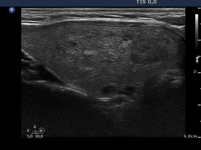
Second examination (second row of images):
Clinical presentation: the patient was referred for a follow-up examination.
Palpation: unchanged.
Functional state: euthyroidism with TSH 2.11 mIU/L.
Ultrasonography. Both the size and the presentation of the nodules remained unchanged.
Comments.
-
Although the larger hyperechogenic granules in the left nodule might be microcalcifications, other properties of the nodule are not suspicious.
-
This case illustrates the difficulty of interpretation of hyperechogenic granules. In such cases the thorough analysis of the video is of much greater help than that of the images. In this particular case we cannot decide how to group these granules.