
Intranodular hyperechogenic figures - case 155
Three years after the previous examination (ultrasonographic picture 3)
|
|

|
|
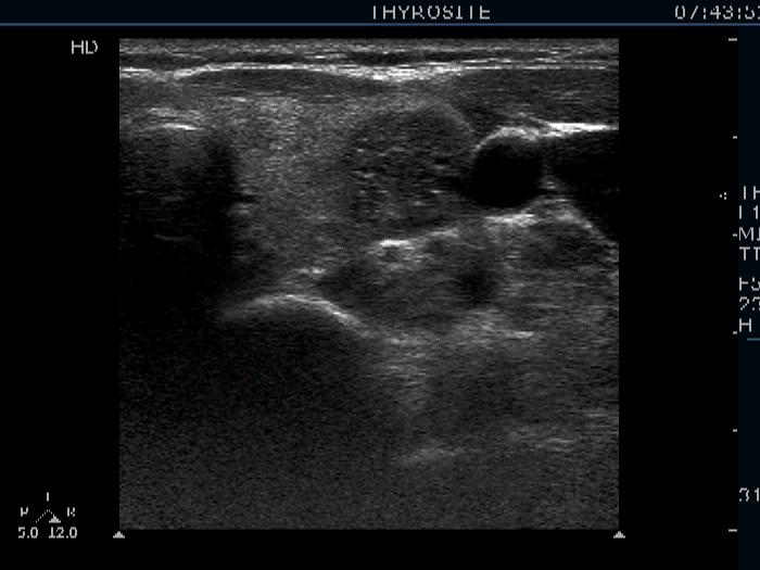
Left lobe, horizontal view. There is a hypoechogenic nodule in the lateral part of the lobe. The nodule contains relatively larger and bright hyperechogenic granules. The presentration of these figures is closer to non-specific granules than to microcalcifications.