
|
|
Differentiation of cystic thyroid lesions - Case 1530doi: 10.24390/thyrocase1530.00
|

|


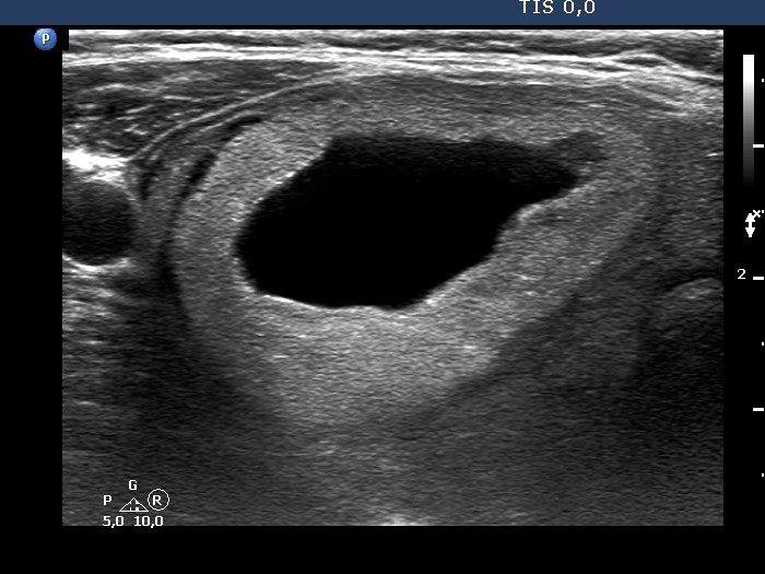

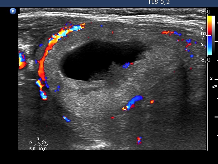


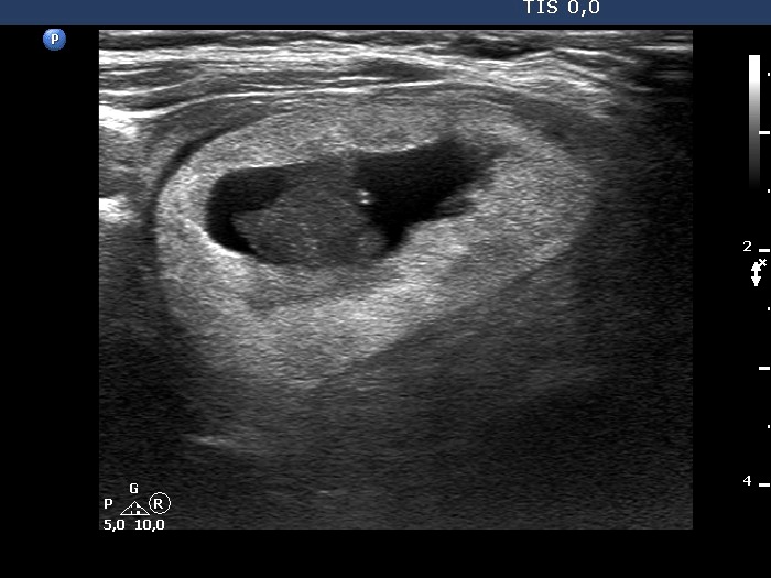

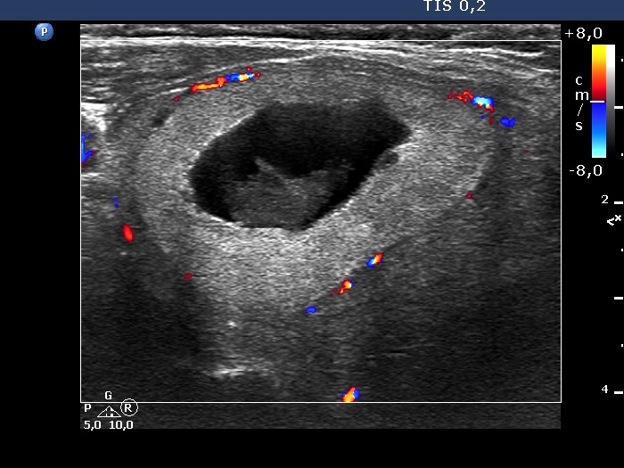
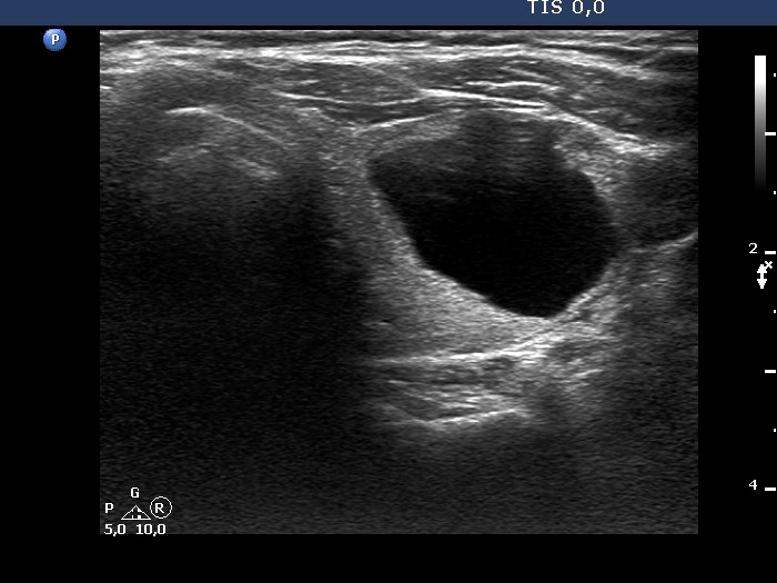

First examination (first row of images)
Clinical presentation. A 68-year-old woman was referred for evaluation of a nodular goiter discovered on routine physical examination.
Palpation: Both lobes had elastic nodule.
Result of blood tests: TSH 4.21 mIU/L, FT4 11.9 pM/L, aTPO 481 U/mL.
Ultrasonography. The thyroid was moderately hypoechogenic. There were two central-type cysts, one in the left lobe and another one in the right lobe. Both had perinodular vascularization.4 mL serous fluid was aspirated. Aspiration cytology resulted in benign cystic lesion.
Suggestion: yearly TSH-determination, ultrasound in three years.
Second examination 3 years later (second row of images)
Clinical presentation. The patient came to routine follow-up visit. She has lost her husband for three months and thereafter diffuse complaints including weight loss, anxiety and sleep disturbance had evolved.
Palpation: unchanged.
Result of blood tests: TSH 5.61 mIU/L.
Ultrasonography. The right cystic nodule has in part refilled. Otherwise, the presentation remained unchanged.Suggestion: TSH determination in 6 months, ultrasound in 3 to 5 years.
Comment. This case illustrates how a spongiform type cyst might evolve from a central-type cystic nodule. See video and longitudinal scans of the right lobe.


