Benign nodular hyperplasia - Case 33. (ultrasonographic picture 4)
|
|

|
|
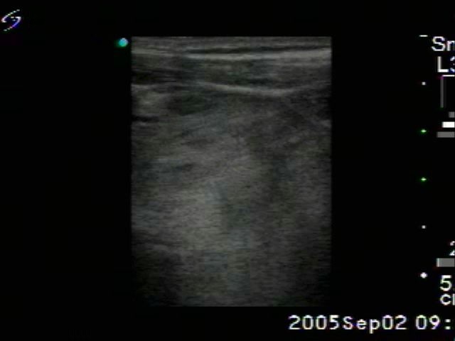
Left lobe, horizontal scan. Here again a greatly enlarged lobe with multiple hyperechogenic nodules.

|
|
|
|
Left lobe, horizontal scan. Here again a greatly enlarged lobe with multiple hyperechogenic nodules.