Benign nodular hyperplasia - Case 41. (ultrasonographic picture 6)
|
|

|
|
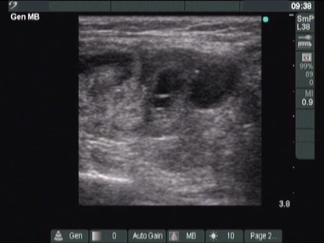
Left lobe, longitudinal scan. There is a hypoechogenic nodule in the lower pole.

|
|
|
|
Left lobe, longitudinal scan. There is a hypoechogenic nodule in the lower pole.