
Benign nodular hyperplasia - Case 4. (ultrasonographic picture 7)
|
|

|
|
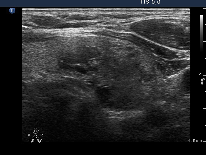
Lower part of the left lobe, horizontal scan. There are two moderately hypoechogenic nodules. The echogenic granules and lines are mostly related to ventral cystic areas, therefore these are back wall figures.