
Benign nodular hyperplasia - Case 14. (cytologic picture 2)
|
|

|

|
|
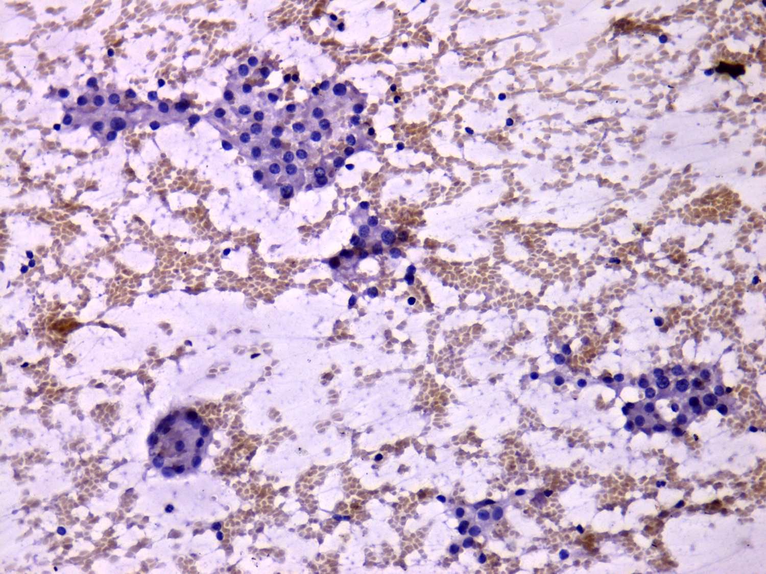
Pap-smear, 200x. Monolayered sheets of microfollicles. The cell group in the left-lower part of the image is specific for a hyperplastic nodule, it is a small cellball.