
|
|
Graves' disease - Case 28.
|
|


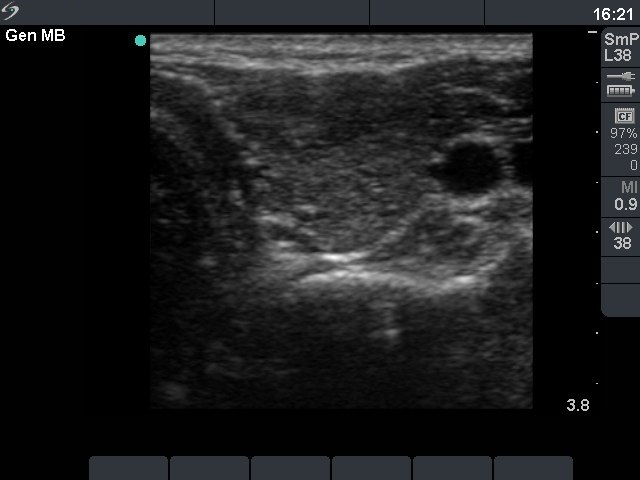
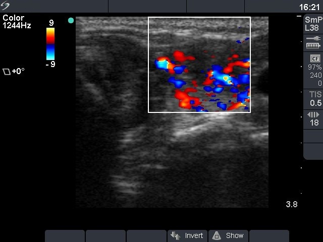


First examination (first row):
Clinical presentation: a 32-year-old woman was referred for an evaluation of typical complaints suggesting hyperthyroidism.
Palpation: no abnormality.
Functional state: hyperthyroidism with TSH 0.001 mIU/L, FT4 52.6 pM/L, FT3 21.6 pM/L.
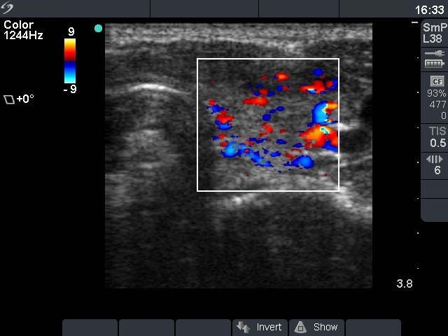
Ultrasonography: the thyroid was moderately hypoechogenic without any nodule. The vascularization was increased.Clinical diagnosis: hyperthyroidism caused by Graves-Basedow's disease.
We administered daily 30 mg methimazole to the patient.
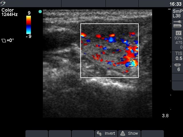
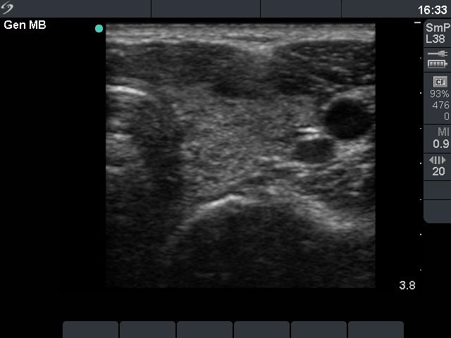
Second examination 5 weeks later (2nd row):
Clinical presentation: most of the original complaints have resolved.
Palpation: no abnormality.
Functional state: subclinical hyperthyroidism with TSH-level 0.02 mIU/L, FT4 12.1 pM/L, FT3 4.08 pM/L.
Ultrasonography: the sonographic pattern did not change over 5 weeks.
Clinical diagnosis: treated hyperthyroidism caused by Graves-Basedow's disease in euthyroid state.
The dose of the methimazole was decreased to daily 10 mg.
Ten months after initial examination (3rd row):
Clinical and laboratory data: the patient was well and euthyroid on daily 5 mg methimazole and 50 microgram levo-tiroxine.
Ultrasonography: the thyroid became echonormal with a 10% echogenicity index. The vascularization was average in the right and decreased in the left lobe.