
Benign nodular hyperplasia - Case 8. (ultrasonographic picture 3)

|

|

|
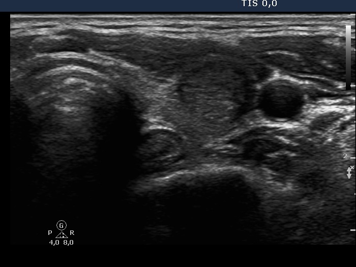
Lower part of the left lobe, horizontal scan. A minimally hypoechogenic, inhomogeneous nodule is presented in this image. The nodule displays halo sign.


