
|
|
Other edifying cases - Case 16: Papillary cancer and Hashimoto's thyroiditis in a lateral aberrant thyroid
|

|



Clinical presentation: a 36-year-old woman was referred for an evaluation of a mass in the right side of the neck known for 4 years. The lesion gradually increased in size and caused complaints when the patient elevated her right arm.
Palpation: a hard lump was palpated above the thyroid bed. There were no nodules in the thyroid on palpation.
Functional state: euthyroidism with TSH 3.61 mIU/L, aTPO 851 U/mL.
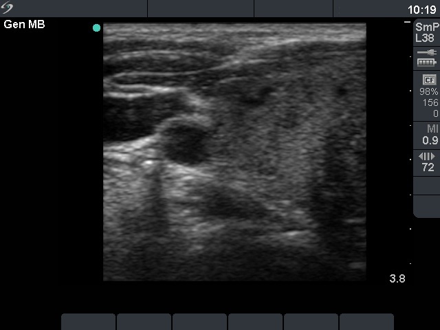
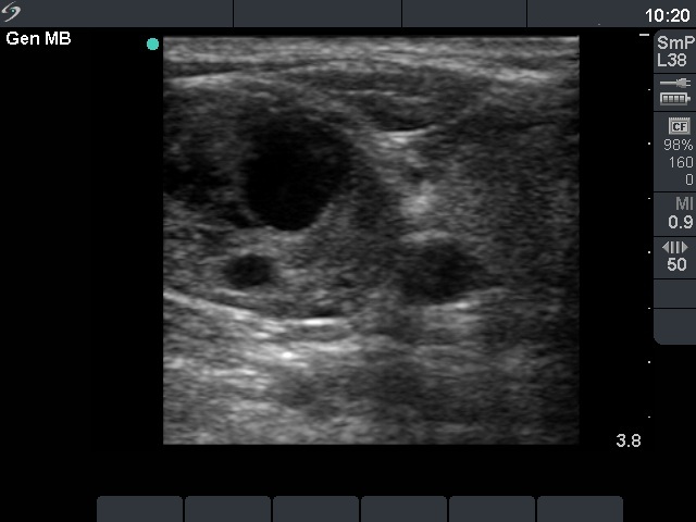
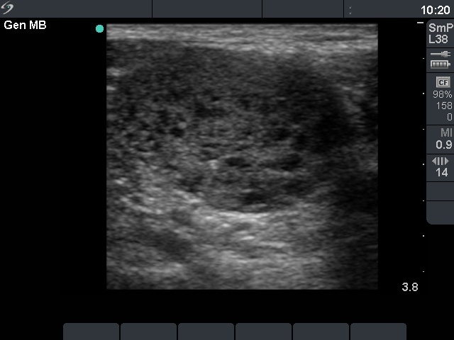
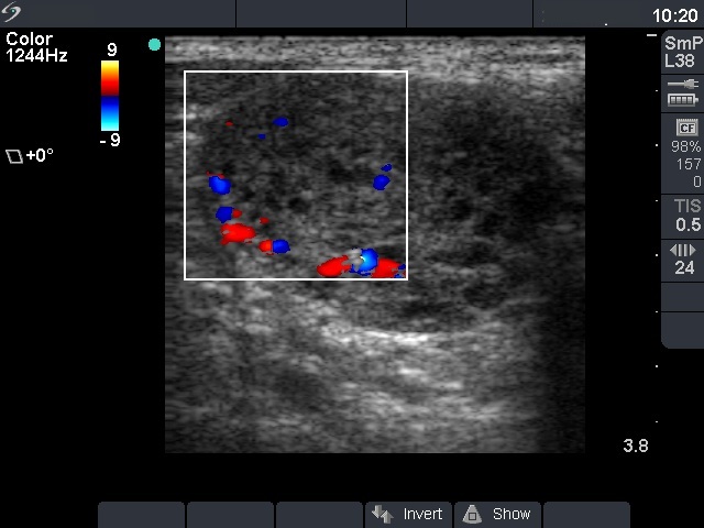
Ultrasonography: both thyroids contained hypoechogenic areas with an echogenicity index of 30%. The palpable lesion was above and lateral to the right lobe. It was moderately hypoechogenic with many cystic areas and presented a type 3 vascular pattern.
Cytology: was performed from the lesion, and minimal amount of cystic fluid was aspirated. There were only macrophages on the smear. The cytological diagnosis was not diagnostic puncture.
Preoperative diagnosis: Hashimoto's thyroiditis. Lateral aberrant thyroid tissue.
Surgery was performed because of the size of the lesion.
Histopathology: disclosed papillary thyroid cancer and Hashimoto's thyroiditis according to the aberrant thyroid and Hashimoto's thyroiditis without malignant focus in the thyroid in the usual position.


