
Papillary carcinoma - Case 38. (ultrasonographic picture 6)
|
|

|
|
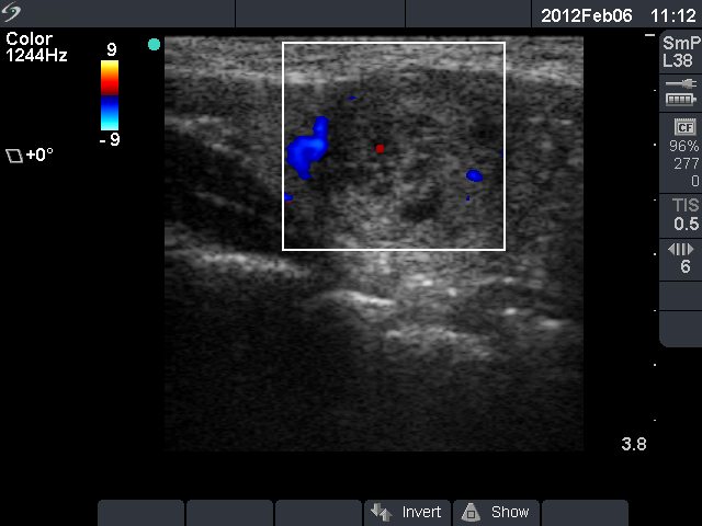
Left lobe, longitudinal scan, color Doppler mode. There is a vessel close to the upper pole of the nodule. Type 1 vascular pattern, i.e. neither perinodular nor intranodular blood flow is detected.