
|
|
Oxyphilic adenoma - Case 6.
|
|


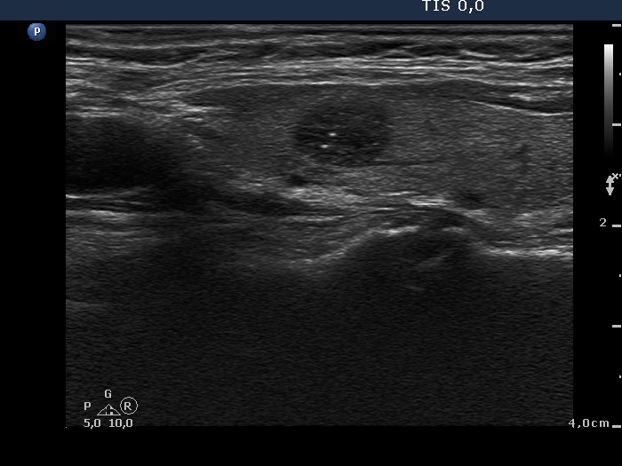


Clinical presentation: a 64-year-old woman was referred for a follow-up examination. Aspiration cytology was performed for 10-ys from a lesion with a maximal diamter of 8 mm-s and resulted in benign lesion. She had no complaints.
Palpation: a small, not firm nodule in the right lobe.
Functional state: euthyroidism with TSH 0.73 mIU/L.
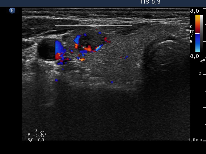
Ultrasonography: revealed a hypoechogenic nodule with microcalcifications in the ventro-lateral part of the right thyroid. The nodule did not present a halo sign while did a combined type 2 and type 3 vascular pattern.
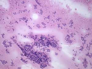
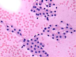
Cytological report: suspicion of a Hürthle-cell tumor.
Histopathology: disclosed Hürthle-cell adenoma.
Comment. The cytological pattern itself might correspond to either a hyperplastic nodule or an oxyphilic adenoma.
.