
|
|
Follicular adenoma - Case 19.
|
|


Clinical data: a 50-year-old woman was referred for a follow-up examination. She was examined for years and the cytology resulted in benign follicular proliferation.
Palpation: the left thyroid was enlarged. The presence of a nodule was equivocal.
Hormonal examination indicated euthyroidism with TSH 1.58 mIU/L.
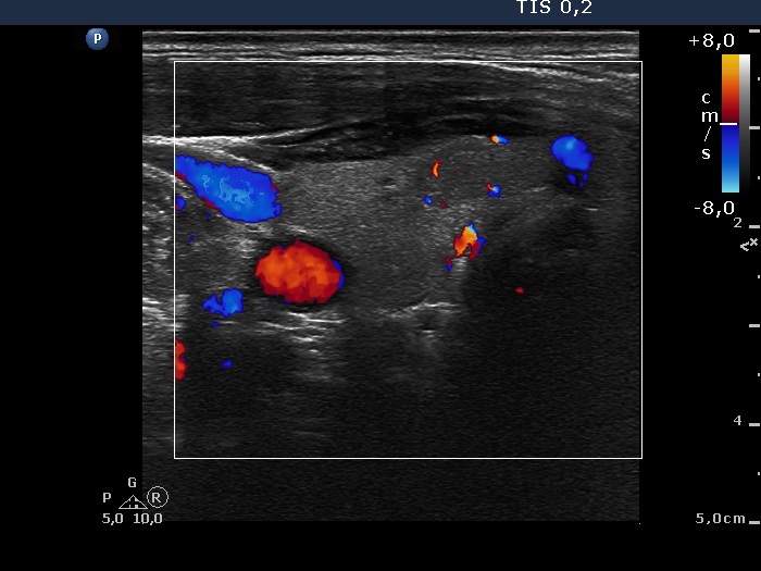
Ultrasonography: the thyroids were echonormal. There was a small hypoechogenic lesion in the ventral part of the right lobe, while a large hyperechogenic nodule presenting a halo sign and perinodular blood flow in the left thyroid. The size of the nodule increased from 25x19x27 mm to 30x27x33 mm (width, depth, length, respectively).
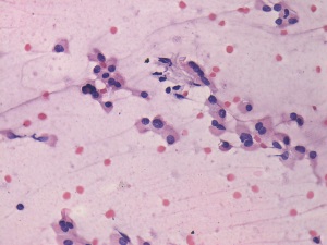
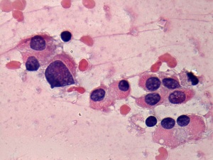
Cytology resulted in suspicion of malignancy, either a follicular variant of papillary cancer or a follicular tumor.
Histopathology disclosed follicular adenoma.
Comment even the unusually high frequency of intranuclear holes has to raise the possibility that these are not pathognomic inclusions. In fact most of these are only artifacts i.e. projections of vacuoles of extranuclear origin.