
Papillary carcinoma - Case 46. (ultrasonographic picture 3)
|
|

|
|
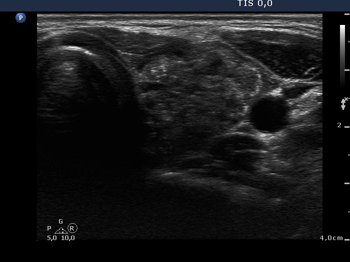
Left lobe, horizontal scan. Almost the entire lobe is consisted of a moderately hypoechogenic, inhomogeneous nodule with numerous bright hyperechogenic foci.