
|
|
Intranodular hyperechogenic figures - case 608doi: 10.24390/thyrocase608.00
|
|


Clinical presentation: A 37-year-old woman came to a follow-up examination. First we met her seven years ago during her pregnancy when a large nodular area was diagnosed. In the subsequent years we three-times aspirated significant amount of cystic fluid. We agreed that surgery will be required and the patient wished to postpone the operation until her child grows a bit older. She noticed an increase in nodule size in the last couple of weeks which caused difficulties in swallowing.
Palpation: a large not firm nodule in the middle portion of the thyroid.
Functional state: euthyroidism with TSH 0.65 mIU/L.
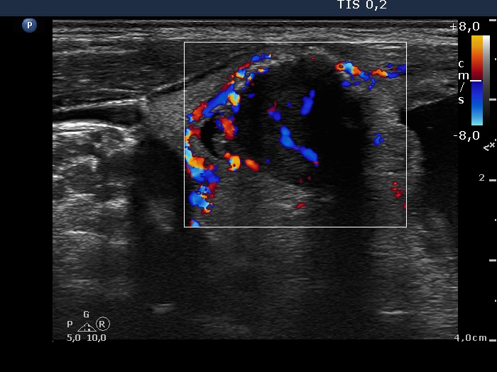
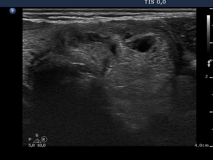
Ultrasonography. The thyroid was echonormal. There was a large mixed nodule having hypoechogenic and echonormal solid areas. The latter presented non-specific hyperechogenic granules and large, irregular hyperechogenic figures, both granules and lines. These correspond to thickened connective tissue. There were several moderately hypoechogenic lesions in the left lobe.
10.5 mL serous fluid was removed than the hypoechogenic solid part was aspirated. Cytology resulted in cystic degeneration.
Surgery was performed. Histopathology disclosed a Hürthle-cell adenoma.
Comments. The amorphous hyperechogenic figure might be connective tissue. It is worth note that one of these thick figures had an incomplete dorsal acoustic shadow.