
Intranodular hyperechogenic figures - case 608 (ultrasonographic picture 3)
doi: 10.24390/thyrocase608.03
|
|

|
|
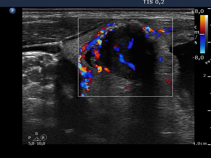
Right lobe, horizontal scan, color Doppler mode. The hypoechogenic area has both perilesional and intranodular blood flow.