
|
|
Papillary carcinoma - Case 73.
|
|


Clinical data: a 64-year-old woman was operated on a sigma carcinoma. On PET-CT scan a positive thyroid nodule and a lymph node in the left supraclavicular region were found.
Palpation: there was a hard nodule in the left thyroid.
Functional state: euthyroidism with TSH 1.90 mIU/L.
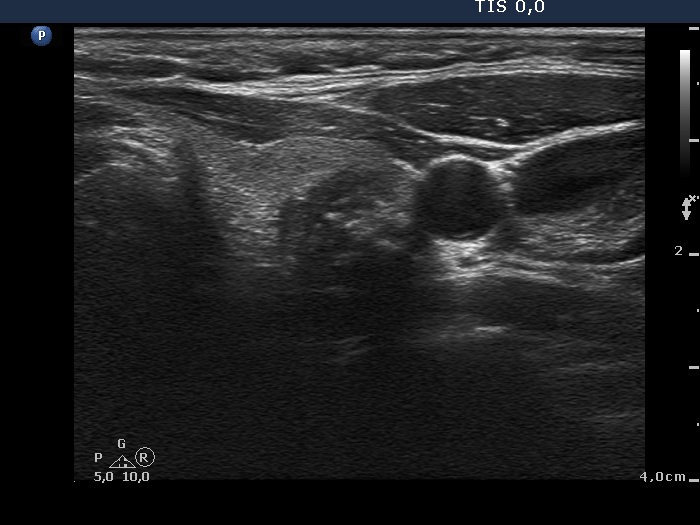
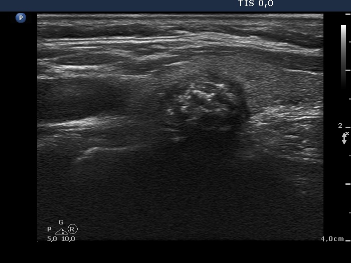
Ultrasonography. The thyroids were echonormal. There were two hypoechogenic nodules, one in the right and another one in the left lobe. The former was regular in shape and presented a type 1 vascular pattern, while the latter presented coarse and microcalcifications. Enlarged lymph nodes without a regular hilum were detected in the left submandibular and supraclavicular regions.
Cytology of the nodule in the left lobe and that of one lymph node resulted in papillary carcinoma.
Total thyroidectomy was performed. Histopathology disclosed papillary carcinoma in the left lobe metastatisizing to the ipsilateral lymph nodes.
Comments.
-
It is worth to compare the benign nodule in the right with the malignant one in the left lobe. The former is a typical benign appearing nodule while the latter presents three different intranodular hyperechogenic figures.
-
There are two different patterns on the same smear. The cells come from benign and from malignant part of the nodule in the left lobe.