Benign nodular hyperplasia - Case 9. (ultrasonographic picture 2)
|
|

|
|
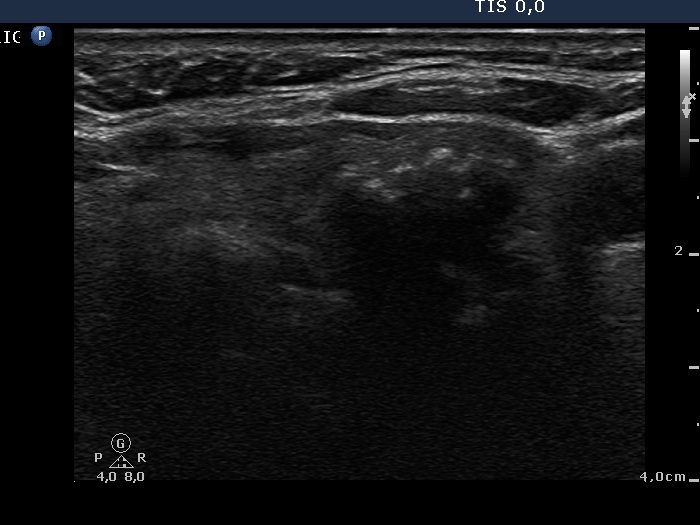
Left lobe, horizontal scan. There is a nodule whic has coarse calcification in its ventral part..

|
|
|
|
Left lobe, horizontal scan. There is a nodule whic has coarse calcification in its ventral part..