
|
|
Intranodular hyperechogenic figures - case 662doi: 10.24390/thyrocase662ct.00
|
|


Clinical presentation: a 40-year-old man came to a follow-up visit. He was operated on 9 years ago when a left lobectomy was performed. Histopathology disclosed follicular adenoma. One year before the present examination a multinodular goiter was found in the right lobe. Cytology resulted in benign colloid goiter. He noticed an increase in nodule size.
Palpation: a multinodular goiter in the right lobe.
Functional state: euthyroidism (TSH 1.57 mIU/L).
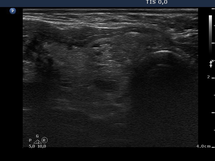
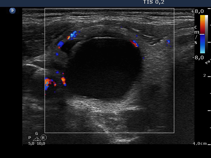
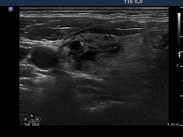
Ultrasonography: the right lobe was echonormal and had multiple lesions. There was a large cystic nodule in the upper part while a nodular area composed of hyperechogenic lesions in the lower part of the lobe. Both nodules contained various hyperechogenic granules.
It became evident after aspirating 7.5 mL brown fluid that the cyst belongs to the spongiform-type cysts.
Cytology resulted in benign cystic degeneration.
We advised a repeat surgery but the patient refused our suggestion. Later he underwent ethanol sclerotherapy.
.