
Differentiation of cystic thyroid lesions - case 723
Four years after the first examination (ultrasonographic picture 1)
doi: 10.24390/thyrocase723.3.01

|

|
 |
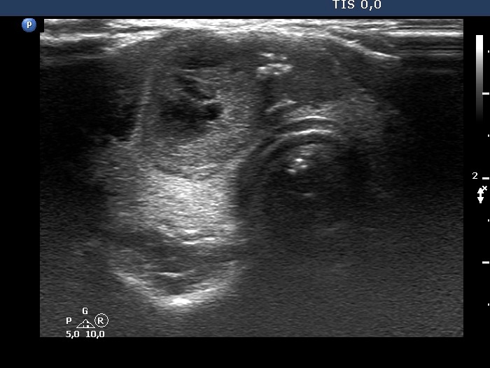
Isthmus, horizontal scan. The nodular area seems to be composed of a larger echonormal and a smaller hypoechogenic lesion, left and right in the image, respectively.


