
Differentiation of cystic thyroid lesions - case 723
Four years after the first examination (ultrasonographic picture 2)
doi: 10.24390/thyrocase723.3.02

|

|
 |
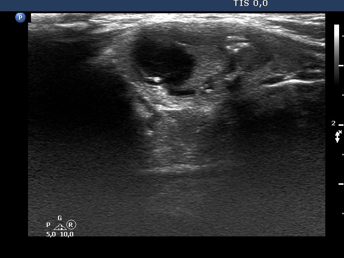
Isthmus, longitudinal scan. TThe cystic content has increased in size. Note the presence of various echogenic figures, 4 of the located in the medial part (right in the image) might correspond to microcalcifications. Nonetheless, similar other figures are clearly related to ventral cystic areas; therefore these four granules are also very likely caused by back wall cystic enhancement.


