
Benign nodular hyperplasia - Case 3. (ultrasonographic picture 2)
|
|

|
|
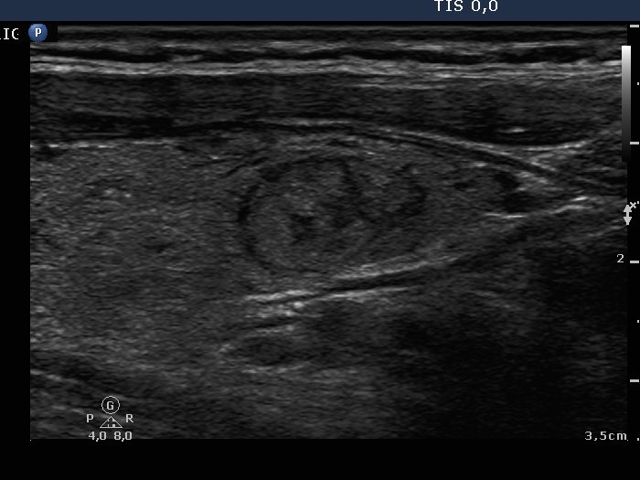
Right lobe, longitudinal scan. Beside the relatively larger lesion in the middle-lower part of the lobe, there are several smaller discrete echo abnormalities.