Benign nodular hyperplasia - Case 3. (ultrasonographic picture 5)
|
|

|
|
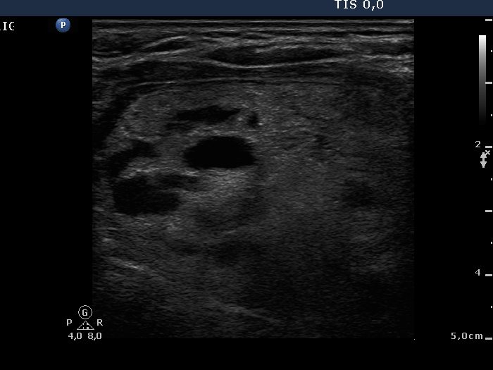
Left lobe, longitudinal scan. A large echonormal-hyperechogenic nodule with cystic degeneration.

|
|
|
|
Left lobe, longitudinal scan. A large echonormal-hyperechogenic nodule with cystic degeneration.