
|
|
The role of complex diagnosis - follow-up of follicular lesions - Case 7.
|
|


First examination (1st and 2nd rows of images)
Clinical data: a 47-year-old woman was referred for an evaluation of a suspected hypothyroidism. She gained in weight 12 kg in the last 2 years.
Palpation: both thyroids were firm. The left thyroid was suspicious containing a nodule.
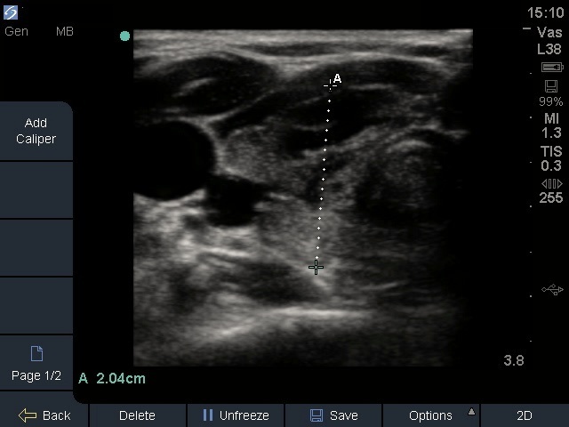
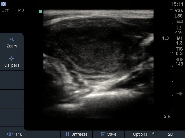
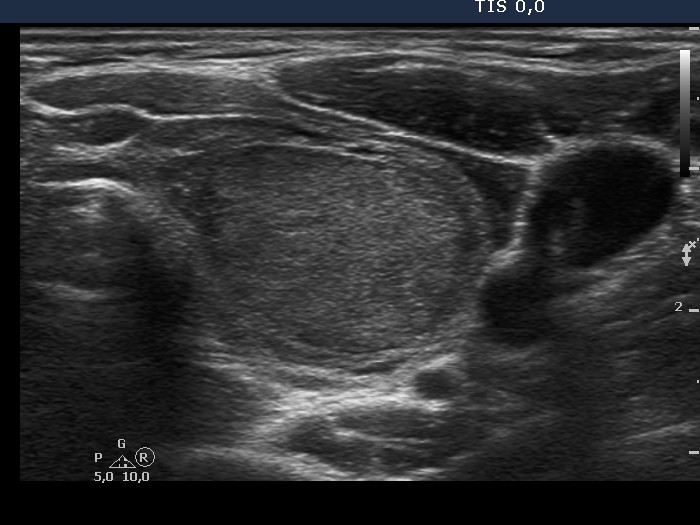
Ultrasonography: the thyroids were echonormal and contained 50% of hypoechogenic areas. There was a minimally hypoechogenic nodule with a halo sign and perinodular blood flow in the left thyroid.
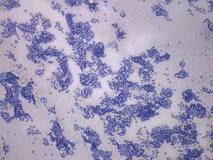
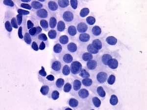
Cytological diagnosis: follicular tumor with less than the average risk of malignancy.
Functional state: euthyroidism with TSH 1.93 mIU/L. Anti-TPO 827 U/mL.
Combined clinical-ultrasound-cytological diagnosis: follicular tumor with not greater than 1% risk of malignancy. Hashimoto's thyroiditis.
We advised follow-up instead of a surgery because the risk of malignancy was estimated not greater than 1%.
Second examination 2 years later (3rd row of images)
Summary of follow-up: the patient underwent yearly ultrasound examination. She had no complaints.
Functional state: euthyroidism with TSH-level 1.44 mIU/L.
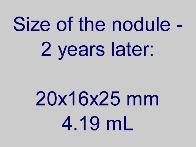
Ultrasonography: the ultrasound presentation of the thyroid was unchanged except for an increase in nodule volume from
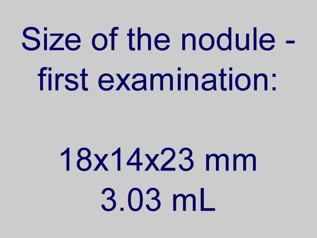
The volume of the nodule was 3.03 and 4.19 mL, at the first examination and at the 2-year follow up, respectively.Surgery was advised.
A left lobectomy was performed and histopathology disclosed follicular adenoma and Hashimoto's thyroiditis in the extranodular part.