
|
|
The role of complex diagnosis - follow-up of follicular lesions - Case 8.
|
|

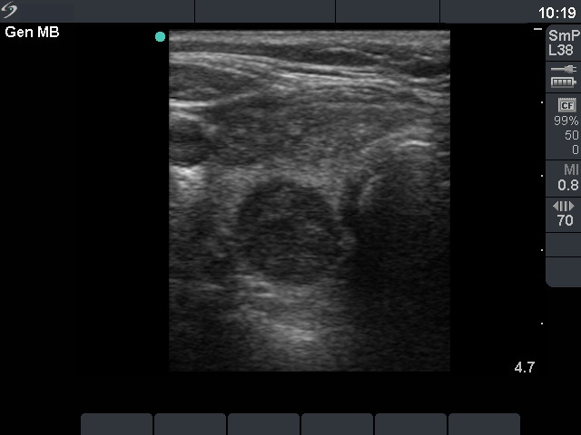
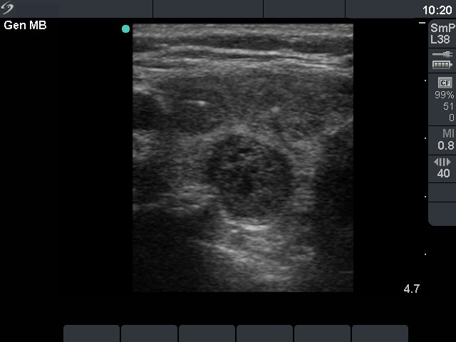

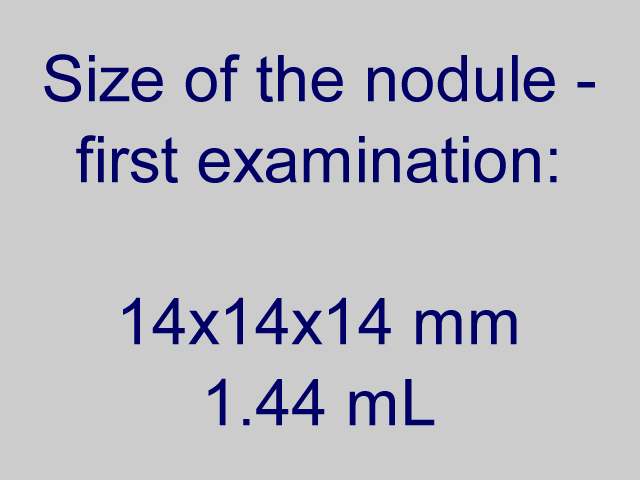
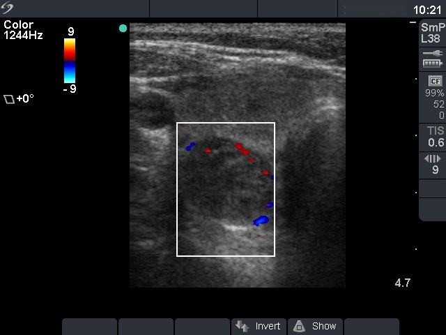
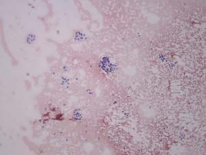
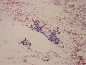
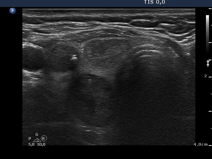

First examination (1st and 2nd rows of images)
Clinical presentation: a 47-year-old woman was referred for evaluation of a multinodular goiter detected on screening. She had no complaints.
Palpation: the right lobe was nodular.
Functional state: euthyroidism with subnormal TSH (TSH 0.18 mIU/L, FT4 18.3 pM/L).
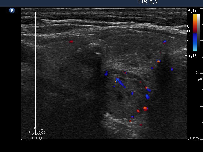
Ultrasonography: the right thyroid contained three nodules, the two ventral lesions were minimally-moderately hypoechogenic, while the dorsal one was hypoechogenic. The latter presented a combined type 2 and type 3 vascular pattern.
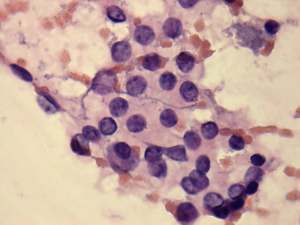
Cytology was performed from the dorsal, hypoechogenic nodule and resulted in benign, follicular proliferation. The risk of a carcinoma was estimated to be less than 1%.
Scintigraphy was performed which disclosed increased uptake in the right lobe. The individual nodules could not be identified.
We advised regular follow-up.
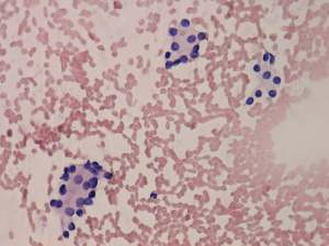
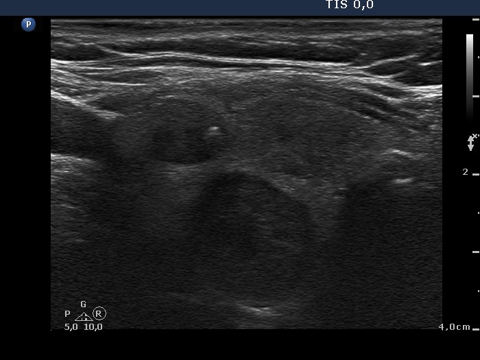
Second examination 2 years later (3rd row of images)
Summary of follow-up: the patient underwent yearly ultrasound examination. She had no complaints.
Functional state: euthyroidism with TSH-level 0.57 mIU/L.
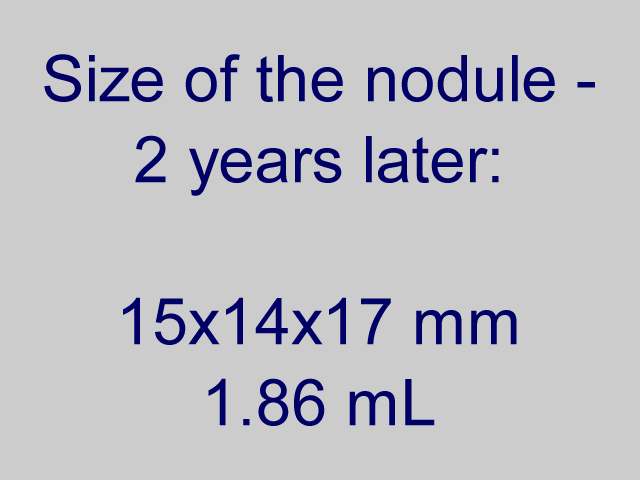
Ultrasonography: the ultrasound presentation of the thyroid was unchanged except for an increase in nodule volume.
Surgery was advised but the patient asked us to go on follow-up examinations.
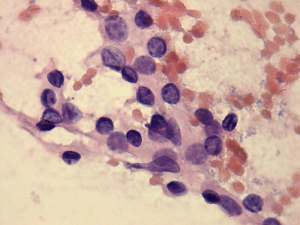
Third examination 4 years after initial examination (4th row of images)
Summary of follow-up: the patient underwent yearly ultrasound examination. She had no complaints.
Functional state: euthyroidism with TSH-level mIU/L.
Ultrasonography: the ultrasound presentation of the thyroid was unchanged except for an increase in nodule volume.
Surgery was advised but the patient asked us to go on follow-up examinations
A left lobectomy was performed and histopathology disclosed follicular adenoma and Hashimoto's thyroiditis in the extranodular part.