
|
|
Chronic lymphocytic thyroiditis - Case 43.
|
|


Clinical presentation: a 57-year-old woman with hypothyroidism known for 13 years. She underwent iris diagnostic investigation where the healer told her that she harbors thyroid malignancy.
Functional state: hypothyroidism (TSH 11.2 mIU/L) on 100 microgram levothyroxine therapy.
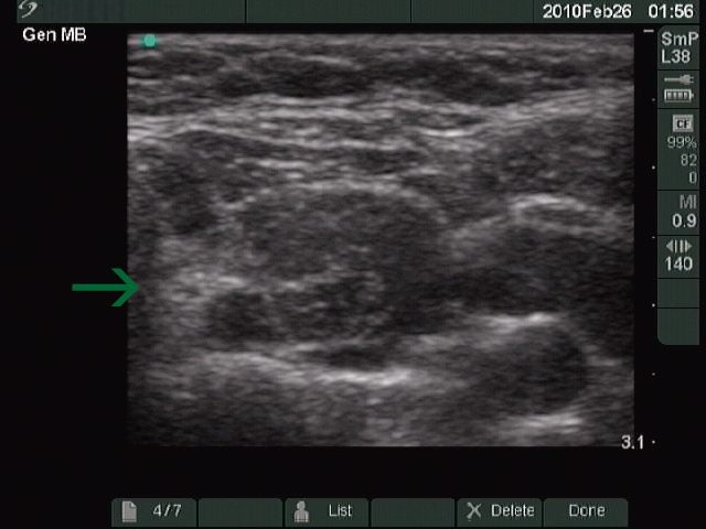
Ultrasonography: multiple hypoechogenic lesions corresponding to Hashimoto's thyroiditis.
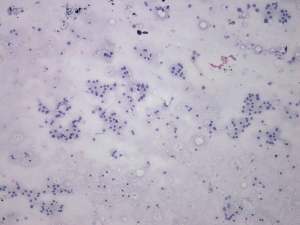
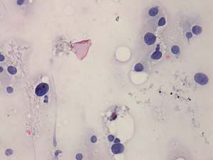
Cytology: no colloid in the background. Almost all thyrocytes exhibit oxyphilic metaplasia. The cells vary in size and shape with several atypical pleomorphic follicular cell. No lymphocytes are present. This pattern fits to Hürthle-cell tumor. However, the lack of prominent nucleoli disfavours the possibility of tumour.
Combined ultrasound-cytological diagnosis: Hashimoto's thyroiditis.
We advised follow-up. The patient went back to the healer who advised surgery.
Histopathology: Hashimoto's thyroiditis without any nodule.
Comment. We frequently gain only oxyphilic cells from Hashimoto's thyroiditis. In several cases, and even in this case too, cytologic picture is identical with a Hürthle-cell tumor. Therefore, it is very important to reevaluate the US picture in those cases where FNAC pattern suggests oxyphilic tumor.