
Benign nodular hyperplasia - Case 44. (ultrasonographic picture 2)
|
|

|
|
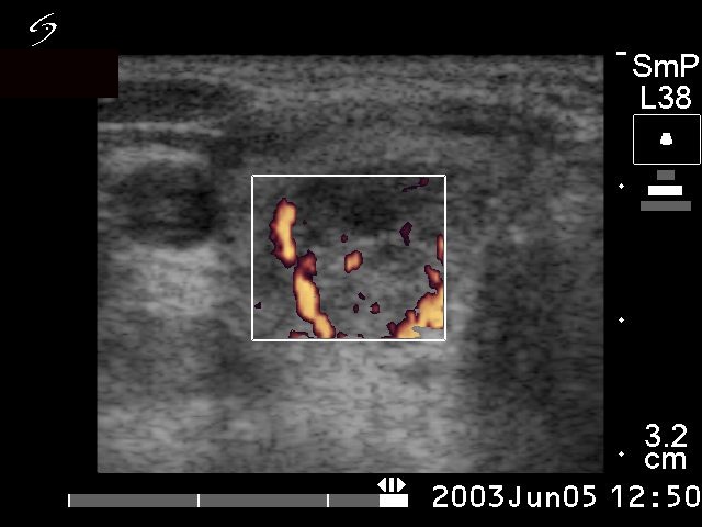
Right lobe, horizontal scan, power Doppler method. Perinodular blood flow with intranodular vascularization. This is the typical vascular pattern of a toxic nodule.