
|
|
Intranodular hyperechogenic figures - case 951
|
|

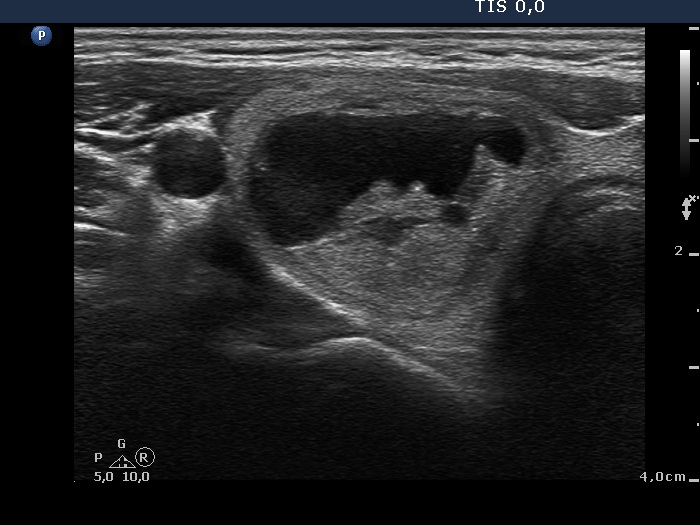


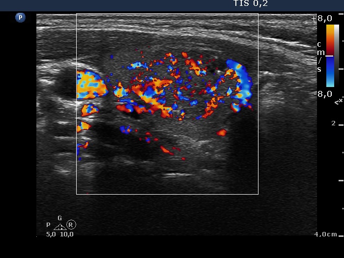

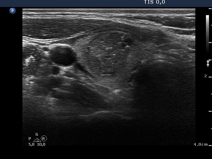


First examination (first row of images):
Clinical presentation: a 30-year-old woman noticed a lump in the right lobe one week ago.
Palpation: a not firm nodule in the right lobe.
Functional state: euthyroidism with TSH 0.83 mIU/L, FT4 14.1 pM/L.
Ultrasonography. The thyroid was echonormal. There were two nodules in the right lobe, the upper one was echonormal-cystic presenting halo sign and perinodular blood flow. It seemed a peripheral-type cyst. The lower was a suspicious nodule: this was hypoechogenic, contained numerous microcalcifications, had a blurred border and lobulated margins and showed an irregularly increased vascularization.
3.5 mL brown fluid was aspirated. Cytology resulted in cystic degeneration and non-diagnostic report, upper and lower nodule in the right lobe, respectively.
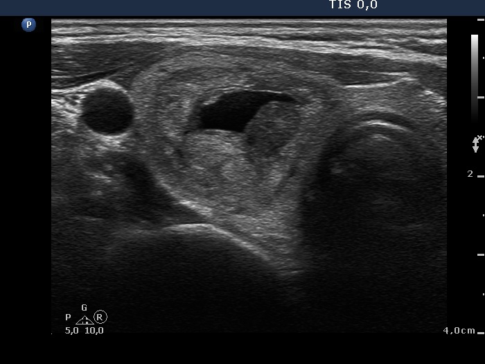
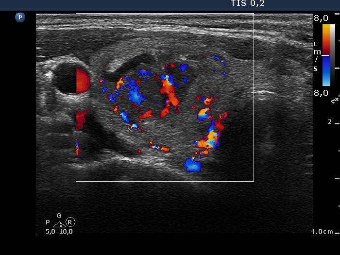
Follow-up examination 2 months later (second row of images):
Clinical presentation: the patient had no complaints.
Palpation: a firm nodule in the right lobe.
Ultrasonography: was basically unchanged. However it became clear that the upper cystic nodule is in fact a central-type cyst.
Cytology of the suspicious nodule was again not diagnostic.
Suggestion: surgery.
A right lobectomy was performed and histopathology disclosed benign hyperplastic nodules.
Comments.
-
This case seems to be an exception: the risk of a papillary carcinoma is above 90% in such nodules. On the other hand, the thorough analysis of the hyperechogenic figures present in the suspicious nodule reveals that these partly belong to non-specific category and partly might be comet-tail artifacts. (It is worth to compare the bright hyperechogenic figures also present in the larger cystic nodule with those found in the lower lesion.)
-
In the event of a non-diagnostic puncture we repeat the cytology after at least 4 weeks. We have found a higher repeatedly non-diagnostic rate if we repeat the aspiration within 2 weeks compared with a reaspiration at least 4 weeks after the first attempt.