
Intranodular hyperechogenic figures - case 951
Follow-up examination 2 months later (ultrasonographic picture 1)
|
|

|
|
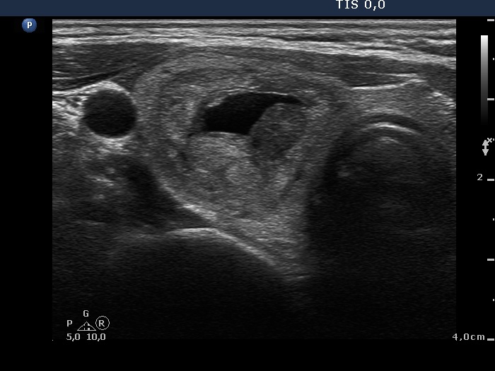
Upper part of the right lobe, horizontal scan. In contrast with the former examination it is clear that this is a central type cyst.