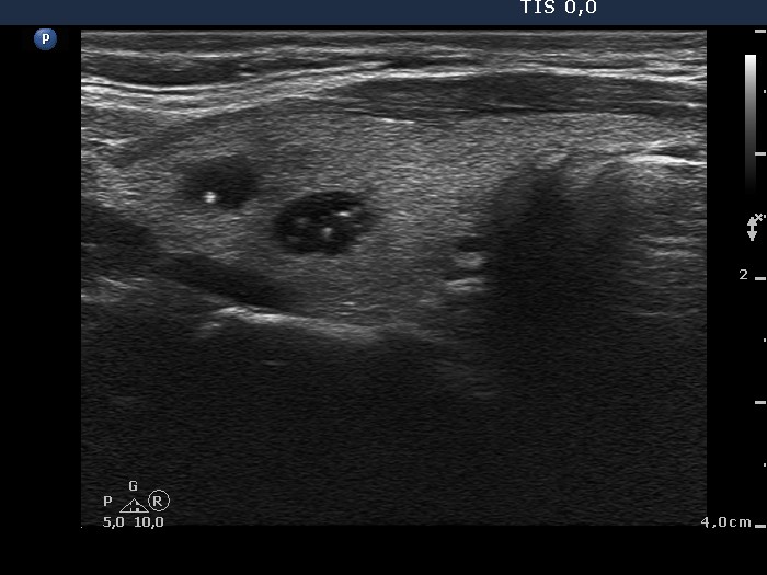
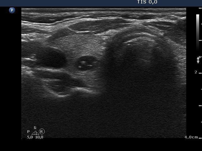
Two small cystic lesions were found in the right lobe, one in the upper and another one in the middle third. Both present hyperechogenic granules. Most of these figures are clearly comet-tail artifacts: they are somewhat blunt and present a tail. The right upper image is very edifying. The granule within the nodule is as bright and sharp as a microcalcification and we cannot see the typical tail in this image. In this case it caused no concern because such a figure within the cystic fluid cannot be a microcalcification - this must be a comet tail artifact, too.