
|
|
Benign nodular hyperplasia - Case 55.
|
|


Clinical presentation: A 49-year-old woman was referred for evaluation of a nodular goiter discovered on screening.
Palpation: no abnormality.
Functional state: euthyroidism (TSH 1.06 mIU/L).
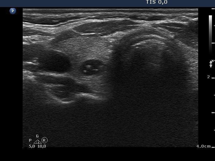
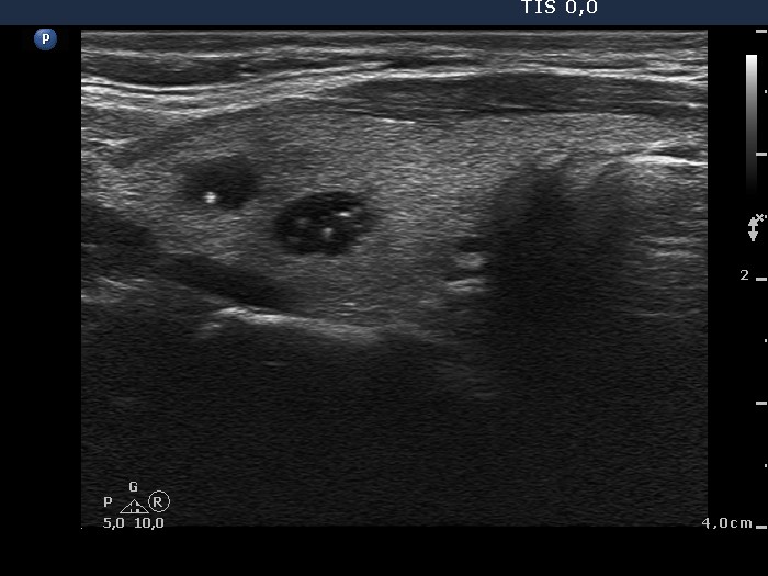
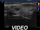
Ultrasonography. The thyroid was echonormal and contained several cystic lesions. The lesions presented numerous comet-tail artifacts. There were two nodules with different echo patterns, a moderately hypoechogenic nodule in the right while a hypoechogenic one in the left thyroid. The former displayed intranodular vascularity while the latter was avascular.
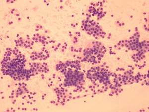
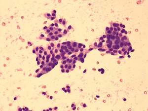
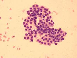
Cytological diagnosis: benign follicular proliferation.
Combined cytological-sonographic diagnosis was benign multinodular goiter, hyperplastic nodules with great probability.
We told the patient that there is a very low risk that a surgical procedure will be required during her lifetime and offered to recheck the size of the small lesions next 3 years later. For unknown reasons the patient was operated on and a bilateral total thyroidectomy was performed.
Histopathology disclosed benign hyperplastic nodules.
Comments.
-
Comet-tail artifacts are protein rich structures and are frequently observed in cystic lesions.
-
The cytological pattern is also remarkable. The lack of colloid, the follicular growth pattern and the presence of monomorphous follicular cells argue for a well-differentiated follicular tumor. On the other hand, the presence of hyperplastic clusters is a weak while the sonographic presentation is a strong argument against a follicular tumor.