|
|||||||||
|
Differential diagnostic of cystic lesions - table 12
|
|||||||||
|
|||||||||
|
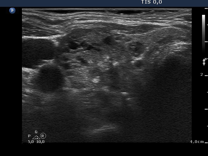
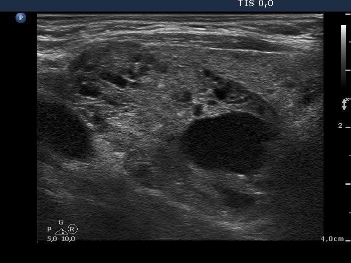
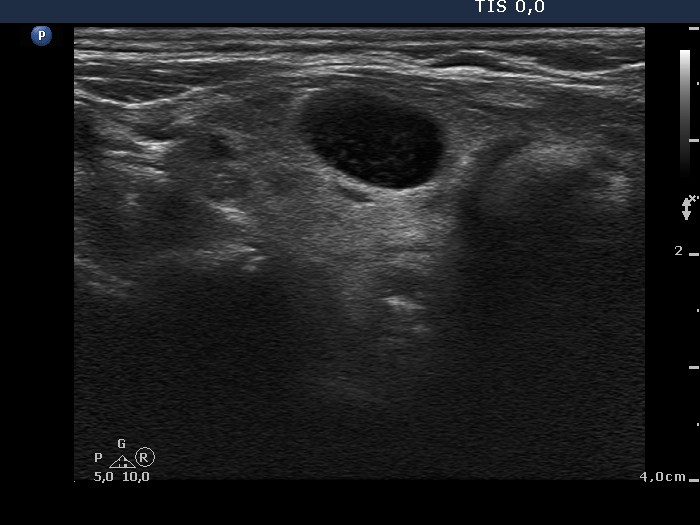
The nodule in the right lobe corresponds to an evolving spongiform cystic lesion. Great proportion of intranodular hyperechogenic figures are non-specific. Nevertheless the presence of microcalcification cannot be fully excluded. The cyst in the left lobe is probably a peripheral type lesion because of the presence of solid part in the lateral part (see upper image). |
|||||||||