|
|||||||||||||||||
|
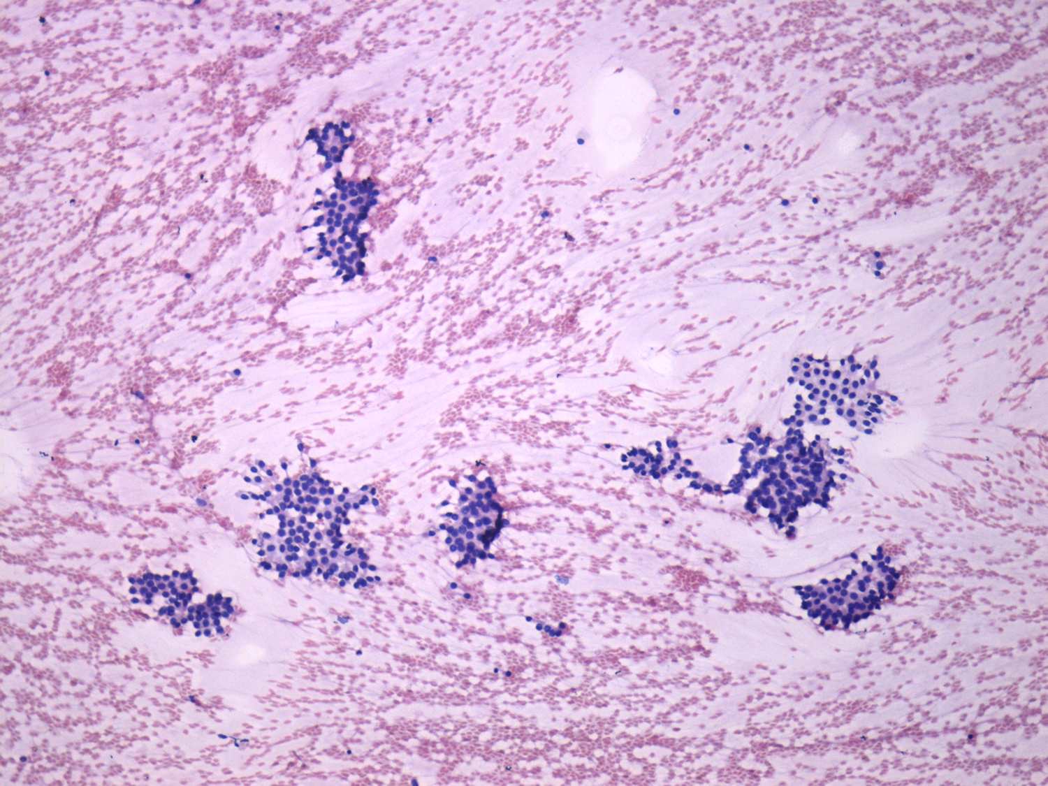
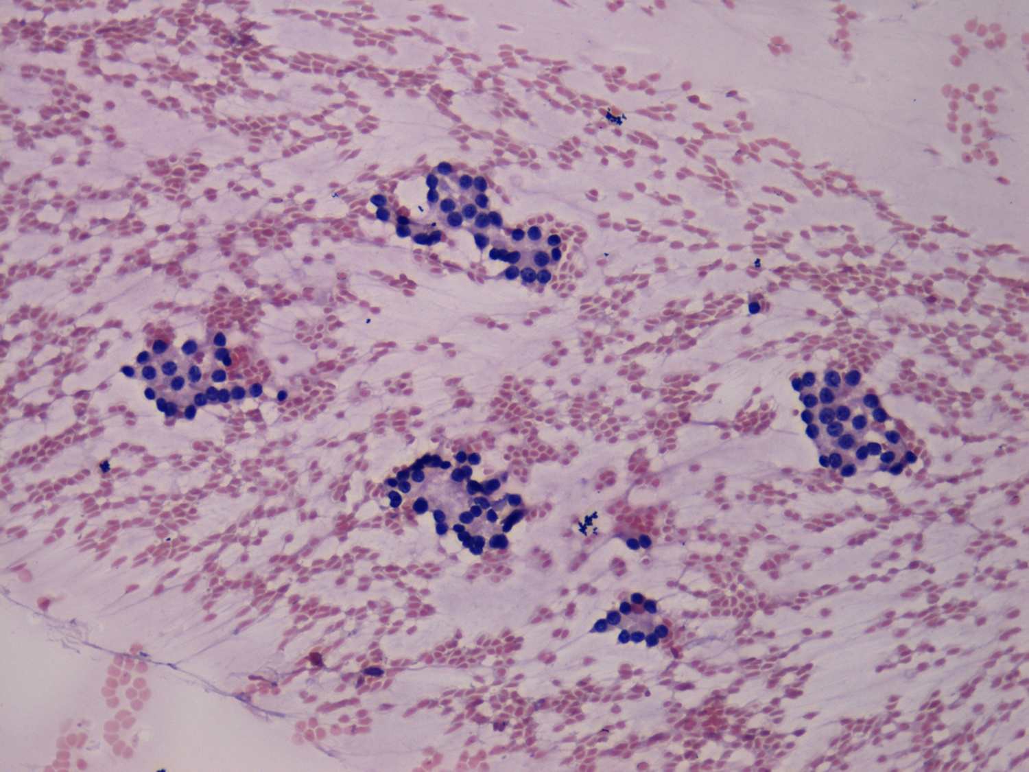
Differential diagnostics of follicular lesions - table 4
|
|||||||||||||||||
|
|||||||||||||||||
|
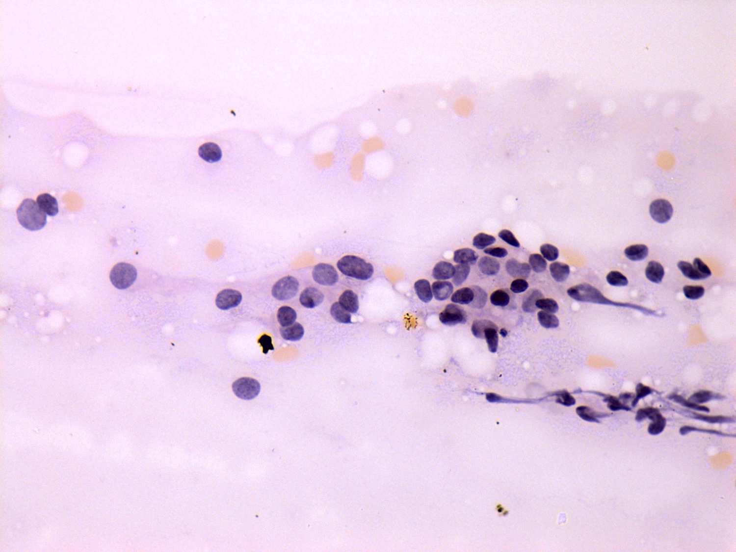
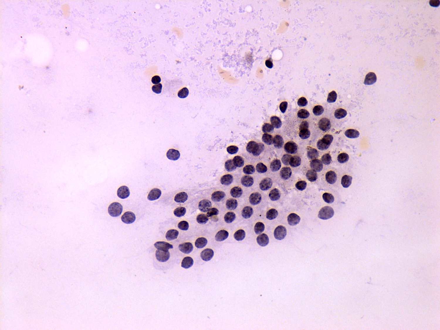
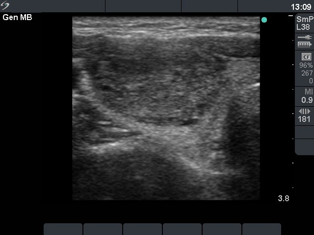
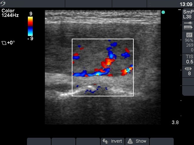
It is worth to compare the structure of the monolayered sheets in the two cases (last row of images). In the event of follicular adenoma, this cluster is composed of microfollicles: almost every nuclei are part of a microfollicle. In hyperplastic nodule we can also identify microfollicles, however most nuclei are hard to fit as a component of a microfollicle. Nevertheless, this difference between the two entities is not qualitative but quantitative. In the last but one image the presentation of monolayered sheet of follicular adenoma is almost identical to that of the hyperplastic nodule presented in the last image. The ultrasound added little to the differential diagnostic of the left patient. Halo sign was not present while the lesion displayed signs of a perinodular flow. It means that we could not exclude the possibility of a follicular tumor on the ultrasound pattern. On the other hand, the ultrasound presentation is almost pathognomic in the left case: a large and solitary nodule displays both halo sign and perinodular blood flow. The combination of this four features means that the likelihood of a follicular tumor exceeds 95%. |
|||||||||||||||||