|
|||||||||||||||||
|
Differential diagnostics of follicular lesions - table 5
|
|||||||||||||||||
|
|||||||||||||||||
|
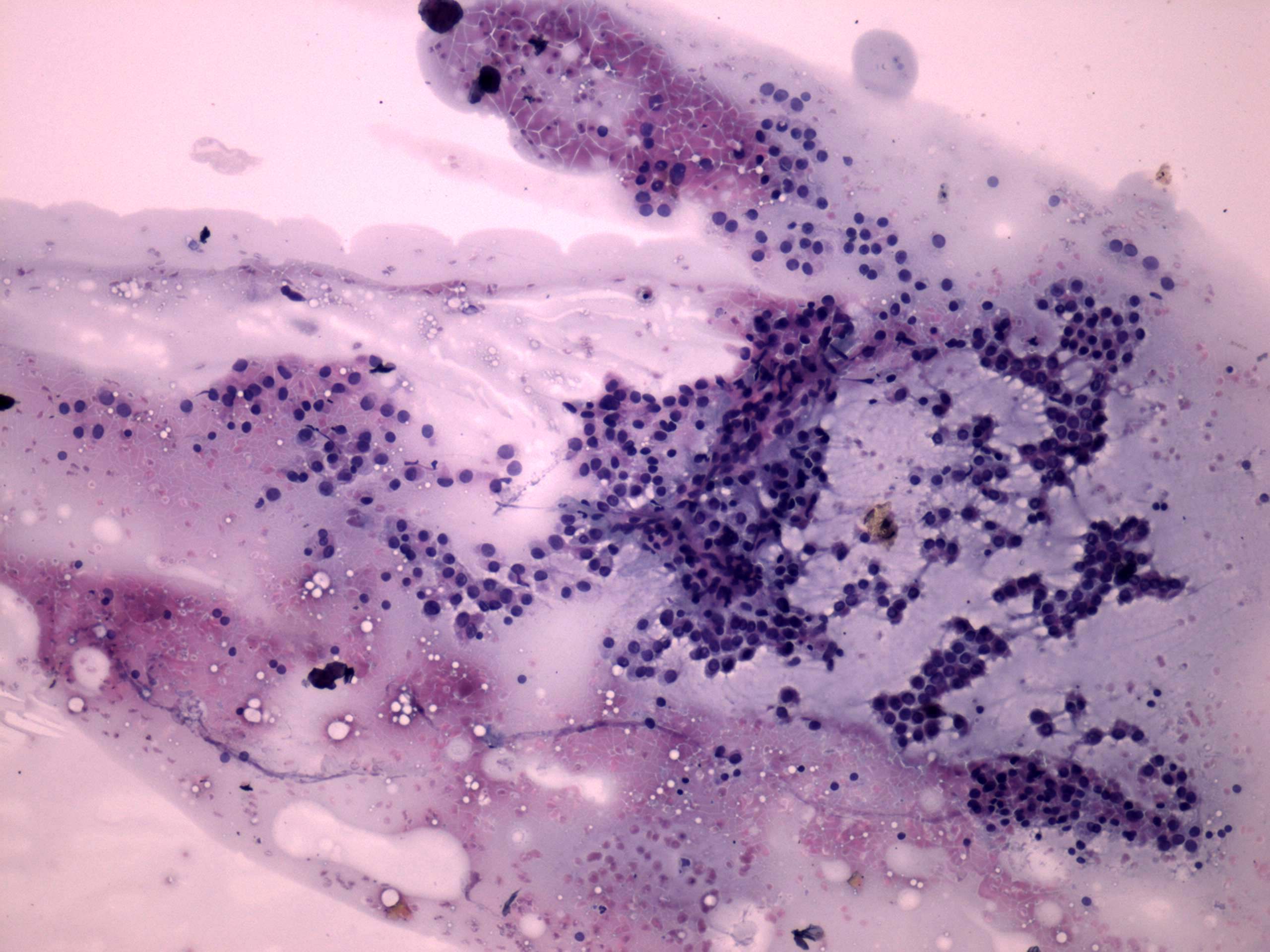
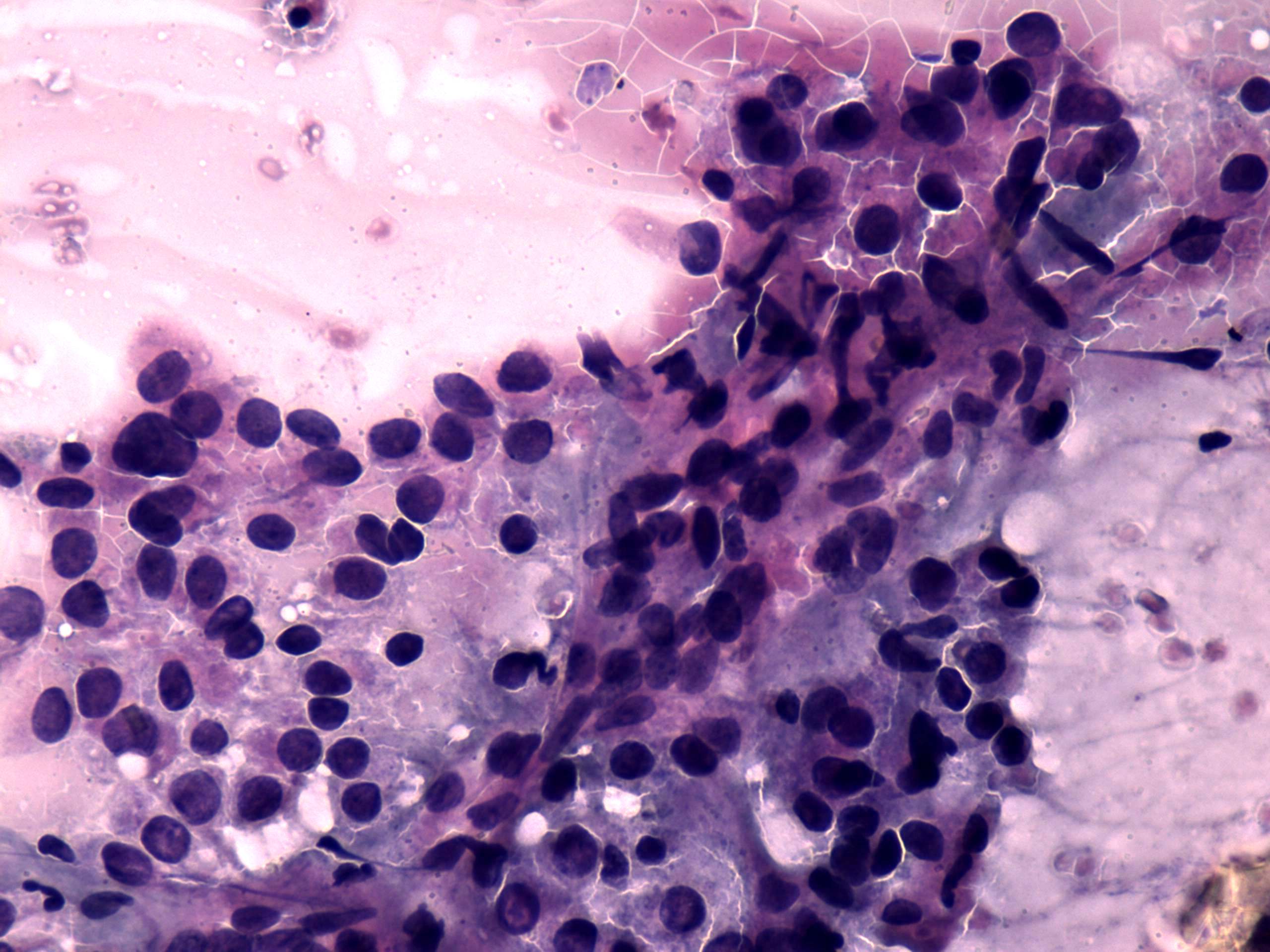
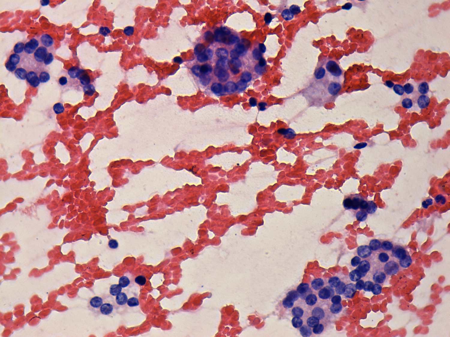
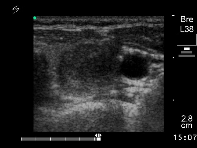
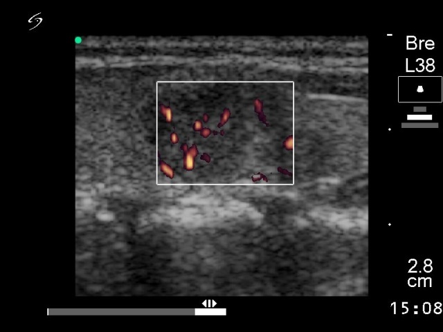
The two cases differ mainly in the presence of colloid and in the proportion of microfollicles. It is worth to compare the large sheet of cells in the first row of images. The individual microfollicles are more easy to recognize in the tumorous case than in the non-neoplastic one. The ultrasound presentations are characteristic in both cases. The hyperplastic nodule present neither a halo nor a perinodular blood flow which decreases the likelihood of a follicular tumor to less than 5%. The nodule in the left case was solitary and presented both perinodular blood flow and halo. |
|||||||||||||||||