
|
|
Benign nodular hyperplasia - Case 28.
|
|


Clinical presentation: a 52-year-old woman with a thyroid nodule known for years. Her nodule increased in size and the patient was informed that her thyroid causes her difficulties swallowing for years. The patient was known for having reflux disease.
Palpation: a firm nodule in the left lobe.
Functional state: euthyroidism (TSH 0.21 mIU/L, FT4 10.1 pM/L).
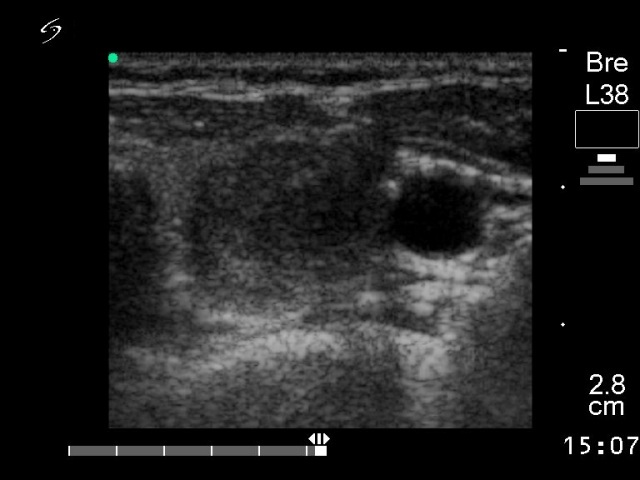
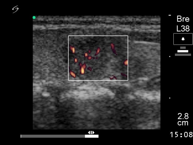
Ultrasonography: a multinodular goiter with a moderately hypoechogenic nodule with increased intranodular vascularity in the left lobe.
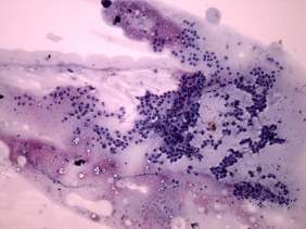
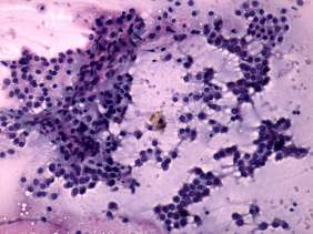
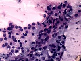
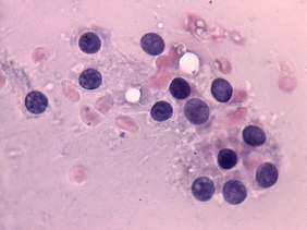
Combined cytological-sonographic diagnosis: benign colloid goiter.
We advised regular follow-up examinations, because the size of the thyroid excluded the possibility of dysphagia. The patient wished to be operated.
Histopathology: benign hyperplastic nodule. Chronic lymphocytic thyroiditis.
Comment: the cytological pattern is not far from atypia of an unknown significance. However, the presence of colloid, the lack of prominent nucleoli are arguments against an oxyphilic tumor. Moreover, if we take the ultrasound into account (i.e. the lack of sonographic signs of a capsule), the oxypilic variant of a follicular tumor can be excluded with great probability.