
Benign nodular hyperplasia - Case 28. (cytologic picture 1)
|
|

|

|
|
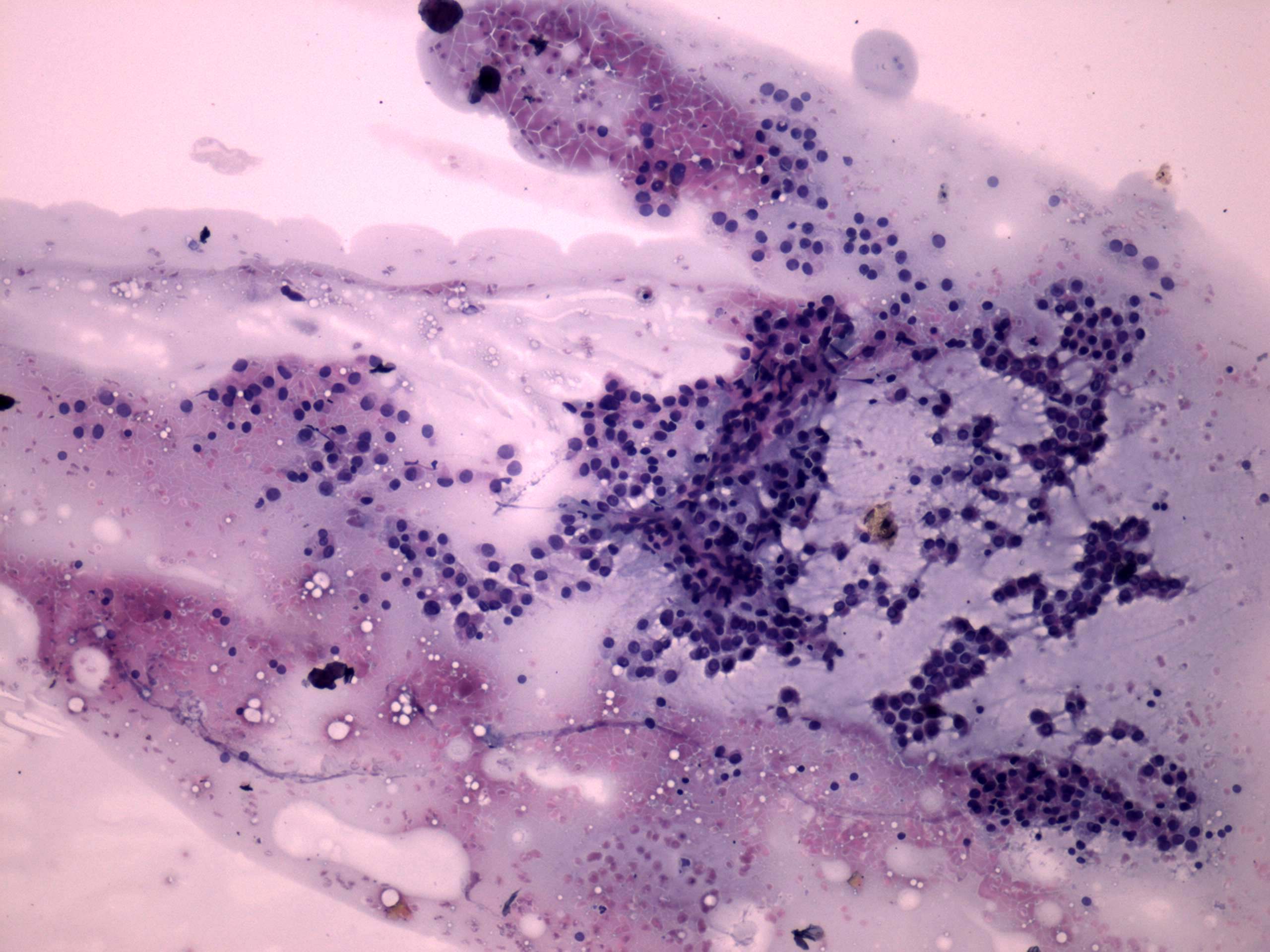
Pap-smear, 100x. Diffuse colloid precipitate in the background. Thyrocytes are arranged predominantly in microfollicles and dissociated.