Benign nodular hyperplasia - Case 28. (ultrasonographic picture 1)

|
|
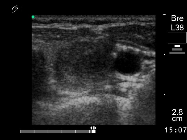
Left lobe, horizontal scan. A moderately hypoechogenic nodule.

|
|
|
Left lobe, horizontal scan. A moderately hypoechogenic nodule.